Translate this page into:
Morphometric analysis of menisci in Nepalese population: A cadaveric study
*Corresponding author: Anuja Jha, Department of Anatomy, Nepal Medical College, Kathmandu, Nepal. dranujanmc@gmail.com
-
Received: ,
Accepted: ,
How to cite this article: Jha A, Pradhan A, Dhungel A, Kharbuja R, Mishra BN. Morphometric analysis of menisci in Nepalese population: A cadaveric study. J Arthrosc Surg Sports Med. 2025;6:37-42. doi: 10.25259/JASSM_41_2024
Abstract
Objectives:
The knee joint’s menisci are wedge-shaped cartilages between the femoral condyles and tibial plateaus. In this study, we investigated and analyzed the diverse morphological variations of knee menisci in human cadavers.
Materials and Methods:
The study was conducted on 50 menisci of embalmed cadaveric knee joints from the Department of Anatomy of Nepal Medical College Teaching Hospital between January 2024 and June 2024. The study examined the morphological variations of the medial and lateral menisci by measuring their circumferences, widths, and thicknesses. Using tools such as a measuring tape, non-elastic threads, metallic pins, and a digital vernier calliper, measurements were taken at three points: Anterior, middle, and posterior thirds.
Results:
Statistical analysis with a one-sample t-test in the Statistical Package for the Social Sciences revealed significant differences between the menisci. The medial meniscus (MM) was predominantly crescent-shaped with a larger inner circumference (P < 0.001), while the lateral meniscus (LM) was more C-shaped with a larger outer circumference (P < 0.001). Width analysis showed the posterior third of the MM as the widest, whereas the middle third was the widest in the LM. Thickness measurements indicated the middle third as the thickest region in both menisci (P < 0.001).
Conclusion:
This study confirms distinct structural differences between the medial and lateral menisci of the knee joint. Understanding these variations enhances diagnostic precision for meniscal injuries, aiding in tailored treatment approaches.
Keywords
Knee joint
Medial meniscus
Lateral meniscus
Morphometric study
Meniscal transplantation
INTRODUCTION
The knee joint’s menisci are wedge-shaped cartilages between the femoral condyles and tibial plateaus. They provide proprioceptive feedback, assist in lubrication, and cushion the underlying bone from the considerable forces generated during extremes of flexion and extension.[1,2] The significance of structural abnormalities and variations within the knee joint has become more pronounced due to advancements in arthroscopy, computed tomography, and magnetic resonance imaging.[3-5] Meniscal injuries are common in various contexts, including sports and everyday activities, and they can have severe consequences.[6] Sports-related joint injuries, particularly in the knees, are common due to high physical activity. It is crucial to have an accurate understanding of the size and shape of the knee meniscus for meniscal transplantation.[7] They play a crucial role in deepening the articular fossae on the tibia, ensuring a more precise alignment for the femoral condyles.[8] The menisci in the knee play a crucial role in providing stability, distributing load, absorbing shock, and maintaining the joint’s health.[9] Injuries to the menisci can cause substantial musculoskeletal problems. Due to their complex structure, treating and repairing meniscal injuries can be challenging for patients and healthcare professionals, including surgeons and physical therapists.[10] The shapes of the menisci are classified into different types based on their morphological variations: Sickle-shaped, U-shaped with sides, V-shaped with sides, crescent-shaped, and C-shaped medial meniscus (MM). The lateral meniscus (LM) was categorized into three types: crescent (semilunar), C-shaped, and Discoid-shaped.[11] The C-shaped MM is wider at the front and covers half of the medial tibial plateau, firmly connected to the joint capsule. In contrast, the circular LM covers 70% of the lateral tibial plateau and is more freely movable.[12] It has strong anterior and posterior attachments but a loose attachment to the lateral joint capsule. This mobility allows greater lateral movement during knee flexion and extension, protecting against lateral meniscal tears.[12,13] The menisci consist of avascular inner and vascular outer zones. Injuries in the avascular zone led to delayed healing, while surgical removal was more effective in the vascular zone. Rising meniscal injuries are linked to increased body mass index and sedentary lifestyles.[14] Detailed knowledge of meniscus size and shape is crucial for successful transplantation in cases of irreparable meniscal damage. Our study aims to determine the morphologic variations in shape, size, width, and thickness of the meniscus among cadavers and also to compare the variations between medial and lateral menisci.
MATERIALS AND METHODS
This descriptive observational, cross-sectional study was conducted on 50 menisci of embalmed cadaveric knee joints at the Department of Anatomy, Nepal Medical College, from January 2024 to June 2024. The research was undertaken following ethical approval from the Institutional Review Committee of Nepal Medical College and Teaching Hospital in Kathmandu.
The inclusion criteria were the cadavers without any morphological abnormalities involved in knee joints, and the exclusion criteria were cadavers involving congenital, prosthetic knee, and pathological abnormalities in their knee joints.
The longitudinal incision was made on each side of the joint capsule, and the patellar ligament and the collateral ligaments were cut transversely. The joint capsule and intraarticular ligaments were cut to expose the menisci, and the condyles were circumferentially separated from their soft-tissue attachments and excised to reveal the tibial plateau. Then, the menisci were exposed, collected, and labeled [Figure 1].

- (a) Collected medial menisci. (b) Collected lateral menisci.
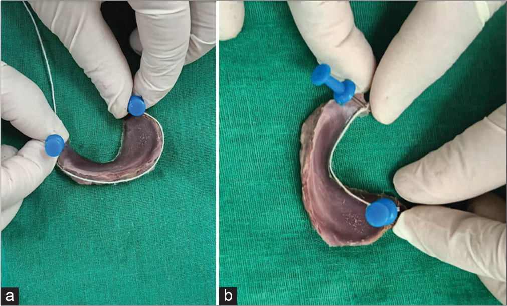
Various shapes of both medial and lateral menisci were observed and categorized based on their morphological variations. The outer and inner peripheral lengths of the menisci were measured using non-elastic cotton threads. The outer length was measured from the most anterior to the most posterior part along the outer margin, while the inner length was measured from the most anterior to the most posterior part along the inner margin. Metallic pins were inserted along the outer margin of the meniscus to support the thread placement [Figure 2]. Subsequently, the length of the thread was straightened and measured using a measuring tape. The same method was followed to measure the inner length of the meniscus.

- (a) Measurement of ‘outer circumference’ of a meniscus using pins and flexible threads. (b) Measurement of ‘inner circumference’ of a meniscus using pins and flexible threads.
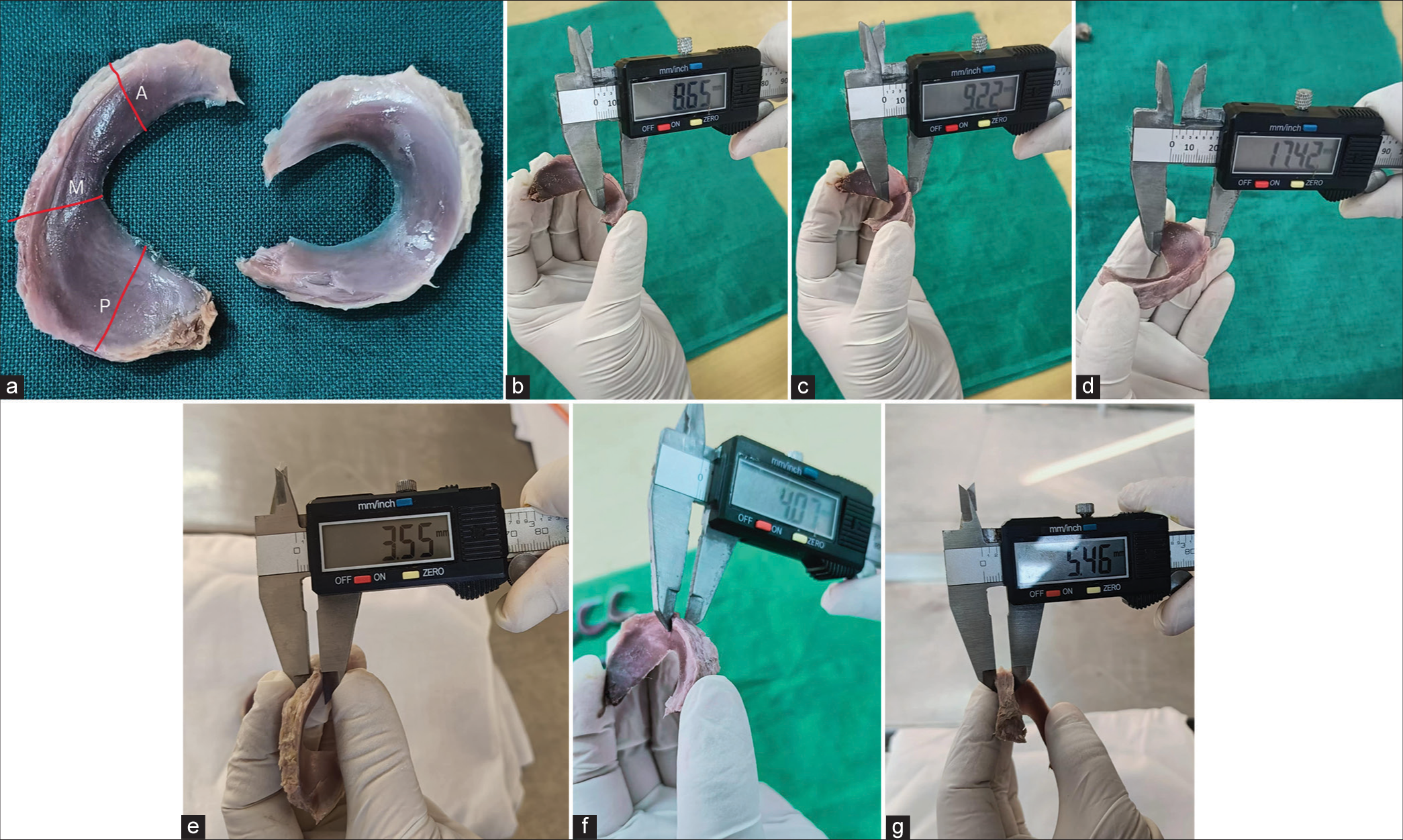
To measure the width and thickness, the menisci were divided into three parts: Anterior (A), Middle (M), and Posterior (P). Maximum width and thickness of each part were measured at three sites: Anterior (A), Middle (M), and Posterior (P). All measurements were recorded using a digital Vernier caliper [Figure 3], and statistical analysis was conducted using the Statistical Package for the Social Sciences version 16.

- (a) Picture representing the three points of morphometric analysis of menisci in the Anterior (A), Middle (M), and Posterior (P) thirds of a meniscus. (b) Meniscal width measurement at anterior 1/3 using digital vernier caliper. (c) Meniscal width measurement at middle 1/3 using digital vernier caliper. (d) Meniscal width measurement at posterior 1/3 using digital vernier caliper. (e) Meniscal thickness measurement at anterior 1/3 using digital vernier caliper. (f) Meniscal thickness measurement at middle 1/3 using digital vernier caliper. (g) Meniscal thickness measurement at posterior 1/3 using digital vernier caliper.
RESULTS
A morphometric study on the knee joint menisci revealed significant differences between the medial and lateral menisci.
Shape
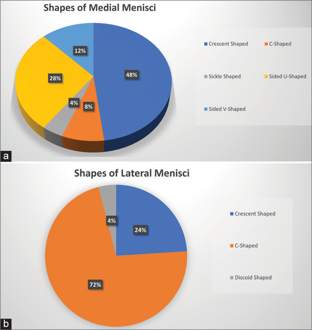
The MM primarily showed crescent shape, while the LM predominantly showed C-shaped structure. The pie diagram showing the incidence of different shapes of medial and lateral menisci is shown in Figure 4.

- Pie diagrams showing the incidence of different shapes of (a) medial menisci and (b) lateral menisci. Crescent shape was most common among medial menisci (48%) whereas C-shape was most common among lateral menisci (72%).
Peripheral length
The analysis reveals no significant difference in the outer circumference between the MM and LM, with a P value of 0.12, suggesting the observed difference is likely due to chance [Table 1]. In contrast, the inner circumference shows a significant difference, with a P < 0.001, indicating that this difference is statistically significant and unlikely to have occurred by random chance. Thus, while the outer circumference does not differ substantially between MM and LM, the inner circumference exhibits a notable difference.
| Observation | Outer circumference | Inner circumference | ||
|---|---|---|---|---|
| MM | LM | MM | MM | |
| Mean | 8.89 | 9.18 | 5.96 | 5.34 |
| Standard deviation | 0.73 | 0.56 | 0.56 | 0.59 |
| Statistical parameter |
t-test: −1.58, P=0.12 |
t-test: 3.81, P<0.001 |
||
MM: Medial meniscus, LM: Lateral meniscus
Thickness
The anterior third of the menisci shows a statistically significant difference in thickness between the medial and lateral menisci (P < 0.0001). The posterior third also shows a statistically significant difference (P < 0.05), whereas the middle third does not show a statistically significant difference in thickness (P > 0.05) [Table 2].
| Observation | Thickness | |||||
|---|---|---|---|---|---|---|
| Anterior third (A) | Middle third (M) | Posterior third (P) | ||||
| MM | LM | MM | LM | MM | LM | |
| Mean | 5.52 | 4.06 | 6.14 | 6.05 | 5.93 | 5.58 |
| Standard deviation | 0.51 | 0.38 | 0.50 | 0.45 | 0.50 | 0.43 |
| Statistical parameter |
t-test 11.48, P<0.0001 |
t-test 0.67, P>0.05 |
t-test 2.65, P<0.05 |
|||
MM: Medial meniscus, LM: Lateral meniscus
Width
The study demonstrates that the MM is significantly narrower than the LM in the anterior (P < 0.001) and middle thirds (P < 0.001) but significantly wider in the posterior third (P < 0.001), revealing consistent regional differences in meniscal width [Table 3].
| Observation | Width | |||||
|---|---|---|---|---|---|---|
| Anterior third (A) | Middle third (M) | Posterior third (P) | ||||
| MM | LM | MM | LM | MM | LM | |
| Mean | 7.66 | 9.53 | 9.84 | 11.48 | 14.90 | 11.14 |
| Standard deviation | 1.19 | 0.41 | 1.39 | 0.66 | 1.25 | 0.61 |
| Statistical parameter |
t-test: −7.42, P<0.001 |
t-test: −5.33, P<0.001 |
t-test: 13.52, P<0.001 |
|||
MM: Medial meniscus, LM: Lateral meniscus
These findings offer comprehensive insights into the morphometric characteristics of the menisci within the knee joint, highlighting their structural variations and potential functional implications, particularly when comparing the medial and lateral aspects.
DISCUSSION
Understanding the variational anatomy of menisci is crucial due to the significant morbidity associated with meniscal injuries and their impact on locomotor function. Meniscal tears are common, particularly in the avascular inner zones, which rarely heal spontaneously. In contrast, peripheral tears can heal satisfactorily if surgically repaired.[15] Therefore, it is essential to conduct studies that document the morphological and morphometric features of knee menisci. Data concerning the morphology of the menisci in this area are limited; therefore, the main objective of this study was to analyze the morphometric variations present in the human meniscus of Nepal.
In the present study, we observed and recorded variations in the morphological features of both lateral and medial menisci. Medial menisci were crescent-shaped (48%), C-shaped (28%), sided V-shaped (12%), sided U-shaped (8%), and sickle-shaped (4%). Discoid shape was not observed in medial menisci, while lateral menisci were C-shaped (72%), crescent-shaped (24%), and discoid-shaped (4%). The findings of our studies are comparable to the findings of Chaware et al.[8] who reported shapes of medial menisci were crescentic-shaped (53%), U-shaped (38.5%), and V-shaped (11.50%), while the lateral menisci were C-shaped (62.5%) and crescentic (37.5%). A study by Murlimanju et al.[16] found that the shapes of the medial menisci are 50% crescent-shaped, 38.9% V-shaped, and 11.1% U-shaped, while the shapes of the lateral menisci are 61.1% C-shaped and 38.9% crescentic. Rao et al.[17] studied 100 knee joints, where they found various shapes of medial and lateral menisci. In medial menisci, 46 were crescentic, 24 were V-shaped, 14 were U-shaped, 11 were C-shaped, and 5 were sickle-shaped. Notably, no discoid-shaped medial menisci were observed. In lateral menisci, 52 were C-shaped, 36 were crescentic, 8 were incomplete discoid, and 4 were complete discoid.
The present study observed a common pattern across various studies, indicating that the MM generally exhibits a greater peripheral length than the LM. This result aligns with findings from various published articles.[8,13,18] However, Hathila et al.[19] presented different findings, suggesting that the peripheral length of the MM and LM can vary [Table 4]. The thickness and width measurements of menisci in this study align with previous findings. It was noted that the anterior third of both the medial and LM was the thinnest, while the middle third was the thickest, consistent with Braz and Silva.[20] The study also found that the MM was narrower at the anterior end and wider at the posterior end, whereas the LM had a consistent width across its length, similar to earlier studies.[8,14,20-23] This variation may be attributed to regional anatomical differences or variations in sample characteristics. These differences highlight the importance of considering geographic and demographic factors when studying meniscal morphology.
| Parameters | Present study | Chaware et al. | Almeida et al. | Braz and Silva | Bhatt et al. | Hathila et al. |
|---|---|---|---|---|---|---|
| Thickness of medial menisci (mm) | ||||||
| Anterior 1/3rd | 5.52±0.85 | 4.45±0.78 | 5.92±1.37 | 6.17±1.68 | 5.82±1.44 | 6.21±0.60 |
| Middle 1/3rd | 6.14±0.94 | 5.50±0.83 | 5.31±1.06 | 6.31±1.73 | 5.64±1.26 | 6.18±0.55 |
| Posterior 1/3rd | 5.93±1.08 | 5.95±1.39 | 5.91±1.13 | 5.18±1.55 | 5.86±1.06 | 6.30±0.42 |
| Width of medial menisci (mm) | ||||||
| Anterior 1/3rd | 7.66±1.25 | 7.05±0.95 | 9.02±1.59 | 7.68±1.36 | 8.78±2.12 | 9.05±0.70 |
| Middle 1/3rd | 9.84±1.28 | 8.12±1.37 | 12.16±2.58 | 9.32±2.24 | 12.08±2.52 | 11.10±0.45 |
| Posterior 1/3rd | 14.90±1.75 | 13.78±2.56 | 17.37±2.22 | 14.96±2.66 | 16.46±2.18 | 15.39±0.80 |
| Thickness of lateral menisci (mm) | ||||||
| Anterior 1/3rd | 4.06±0.93 | 5.41±1.14 | 3.71±1.15 | 4.40±0.83 | 3.70±1.52 | 4.15±0.50 |
| Middle 1/3rd | 6.05±1.02 | 6.55±1.06 | 6.10±1.04 | 6.52±1.81 | 5.78±1.22 | 5.90±0.61 |
| Posterior 1/3rd | 5.58±0.76 | 7.09±1.59 | 5.29±0.78 | 5.46±1.19 | 5.20±0.98 | 5.63±0.60 |
| Width of lateral menisci (mm) | ||||||
| Anterior 1/3rd | 9.53±1.12 | 8.00±2.38 | 11.86±1.81 | 11.32±1.46 | 11.30±1.30 | 11.82±0.81 |
| Middle 1/3rd | 11.48±1.62 | 11.54±3.56 | 11.97±2.56 | 11.16±1.64 | 11.66±1.48 | 12.53±0.72 |
| Posterior 1/3rd | 11.14±1.02 | 12.65±3.46 | 11.44±1.07 | 11.67±1.54 | 11.50±1.34 | 12.03±0.80 |
CONCLUSION
The present study confirms variations in shape, circumference, width, and thickness between the medial and lateral menisci of the knee joint. Understanding these structural variations enhances accuracy in diagnosing meniscal injuries, facilitating tailored treatment approaches. Recognition of the wider posterior third in the MM guides surgical planning for tears in this region, optimizing intervention strategies.
Acknowledgments
We extend our heartfelt gratitude to Prof. Dr. Shaligram Dhungel, Department of Anatomy for his invaluable guidance and support throughout this study. We also wish to express our sincere thanks to all the faculty members of the Department of Anatomy for their continuous support and encouragement.
Author contributions
AJ is the principal investigator and made substantial contributions to conception, design, data acquisition, data analysis to writing of manuscript. AP made substantial contribution to analysis and interpretation of data and to revising of the manuscript. AD and RK made substantial contributions to acquisition of data and to literature review. BNM made substantial contribution to conception and design, literature review, and writing of manuscript. All authors read and approved the final manuscript.
Ethical approval
The research/study was approved by the Institutional Review Board at Nepal Medical College-Institutional Review Committee (NMC-IRC), number 43-080/081, dated January 18, 2024.
Declaration of patient consent
Patient’s consent is not required as there are no patients in this study.
Conflicts of interest
There are no conflicts of interest.
Use of artificial intelligence (AI)-assisted technology for manuscript preparation
The authors confirm that there was no use of artificial intelligence (AI)-assisted technology for assisting in the writing or editing of the manuscript and no images were manipulated using AI.
Financial support and sponsorship
Nil.
References
- Gray’s anatomy: The anatomical basis of clinical practice (40th ed). London: Churchill Livingstone Elsevier; 2008.
- [Google Scholar]
- Anatomic variations of the shape of the menisci: A neonatal cadaver study. Knee Surg Sports Traumatol Arthrosc. 2006;14:975-81.
- [CrossRef] [PubMed] [Google Scholar]
- Clinically oriented anatomy (4th ed). Philadelphia, PA: Lippincott Williams and Wilkins; 1999. p. :6909.
- [Google Scholar]
- Meniscal sizing before allograft: Comparison of three imaging techniques. Knee. 2018;25:841-8.
- [CrossRef] [PubMed] [Google Scholar]
- Meniscus insertion anatomy as a basis for meniscus replacement: A morphological cadaveric study. Arthroscopy. 1995;11:96-103.
- [CrossRef] [PubMed] [Google Scholar]
- The menisci of the knee joint. Anatomical and functional characteristics and a rationale for clinical treatment. J Anat. 1998;193:16178.
- [CrossRef] [PubMed] [Google Scholar]
- Morphological study of the menisci of the knee joint in human cadaver in Jharkhand population. J Family Med Prim Care. 2022;11:4723-9.
- [CrossRef] [PubMed] [Google Scholar]
- A cadaveric study to define morphology and morphometry of human knee menisci in the region of central India. Cureus. 2023;15:e41174.
- [CrossRef] [Google Scholar]
- Morphometric study of menisci of knee joints in adult cadavers. Int J Anat Res. 2016;4:2973-8.
- [CrossRef] [Google Scholar]
- The basic science of human knee menisci: Structure, composition, and function. Sports Health. 2012;4:340-51.
- [CrossRef] [PubMed] [Google Scholar]
- Shapes of menisci of knee joint: A human cadaveric study. Int J Anat Res. 2019;7:6239-43.
- [CrossRef] [Google Scholar]
- Morphometric differences between the medial and lateral meniscus in healthy men-a three-dimensional analysis using magnetic resonance imaging. Cells Tissues Organs. 2012;195:353-64.
- [CrossRef] [PubMed] [Google Scholar]
- Meniscal preservation is important for the knee joint. Indian J Orthop. 2017;51:576-87.
- [CrossRef] [PubMed] [Google Scholar]
- A study on the morphometry of a medial meniscus in the knee joint of human cadavers in the South Indian population. Cureus. 2023;15:e42753.
- [CrossRef] [Google Scholar]
- Preoperative sizing of meniscal allografts in meniscus transplantation. Am J Sports Med. 2000;28:524-33.
- [CrossRef] [PubMed] [Google Scholar]
- Morphometric analysis of the menisci of the knee joint in south Indian human fetuses. J Morphol. 2010;28:1167-71.
- [CrossRef] [Google Scholar]
- Morphometric analysis of the menisci of the knee joint in population of east Godavari region of Andhra Pradesh. J Evol Med Dent Sci. 2014;3:8972-9.
- [CrossRef] [Google Scholar]
- Morphological study of menisci of knee joint in human cadavers. J Anat Soc India. 2017;66:53-4.
- [CrossRef] [Google Scholar]
- Morphological study of menisci of knee joint in human cadavers. J Anat Radiol Surg. 2018;7:AO10.
- [Google Scholar]
- Morphometric study of menisci of the knee joint. Int J Morphol. 2004;22:181-4.
- [CrossRef] [Google Scholar]
- Morphometric study of menisci of knee joint in the west region. Int J Basic Appl Med Sci. 2014;4:95-9.
- [Google Scholar]
- Knee menisci: Structure, function, and management of pathology. Cartilage. 2017;8:99-104.
- [CrossRef] [PubMed] [Google Scholar]


