Translate this page into:
Arthroscopic meniscus transplant using autologous semitendinosus with bone marrow aspirate-derived fibrin clot along with anterior cruciate ligament reconstruction – A case report
*Corresponding author: Himanshu Gupta, Sports Injury Centre, Vardhman Mahavir Medical College and Safdarjung Hospital, New Delhi, India. himanshu. aiims@gmail.com
-
Received: ,
Accepted: ,
How to cite this article: Gupta H, Jhamb JA, Das J, Mishra P. Arthroscopic meniscus transplant using autologous semitendinosus with bone marrow aspirate-derived fibrin clot along with anterior cruciate ligament reconstruction – A case report. J Arthrosc Surg Sports Med. 2025;6:70-3. doi: 10.25259/JASSM_44_2024
Abstract
Subtotal and total meniscectomy is known to advance osteoarthritis in patients. Meniscus transplant using semitendinosus autograft is a viable option in young patients. We have presented the early follow-up, along with technical video, for arthroscopic meniscus transplant with autologous semitendinosus graft with bone marrow aspirate-derived fibrin clot along with anterior cruciate ligament (ACL) reconstruction, in the primary surgery, in a patient of ACL tear along with near-complete loss of medial meniscus due to auto-meniscectomy. The patient showed good functional and radiological outcome at 9 months.
Keywords
Meniscus transplant
Autologous
Semitendinosus
Arthroscopic
INTRODUCTION
Meniscal repair is considered whenever possible in young individuals with meniscus tears. However, in old, neglected injuries or after failure of repair, a subtotal or total meniscectomy has to be performed. Meniscus allograft transplant or replacement is an emerging treatment option with some studies showing good long-term results.[1] However, there are several problems with allografts such as the risk of transplanting foreign tissue, size mismatch, availability, and high-cost. Some recent reports have reported promising outcomes following arthroscopic meniscus transplant using autologous semitendinosus graft.[2,3]
Meniscal transplants have generally been used in cases that had previously undergone a sub-total or total meniscectomy and have become symptomatic[4] with early cartilage changes having set in. Several systematic reviews have concluded that subtotal or total meniscectomies accelerate the process of knee osteoarthritis (OA).[5] We believe that a patient undergoing a sub-total meniscectomy should be offered a meniscus transplant without waiting for them to develop OA.
CASE REPORT
We present a report of a young male with an anterior cruciate ligament (ACL) tear along with near-complete loss of medial meniscus (due to auto-meniscectomy), and focal grade III cartilage lesion in medial femoral condyle. He was managed with ACL reconstruction, and meniscus transplant with autologous semitendinosus graft in the primary sitting. A combination of techniques described by Iida et al. and Rönnblad et al. was used, utilizing bone marrow aspirate (BMA)-derived fibrin clot sandwiched into the graft.[2,3] A description of the technique along with video is also included in the study.
A 36-year-old male, working in the paramilitary force had a history of knee injury 1 year back. Since then, he complained of knee pain accentuated with weight-bearing, with multiple episodes of instability and locking. On examination, Lachman and pivot shift tests were positive and there was medial joint line tenderness. On magnetic resonance imaging (MRI), he was found to have a complete tear of ACL and the medial meniscus involving both roots (with near-total absence of meniscus tissue, suggestive of auto-meniscectomy), focal articular cartilage loss on the medial femoral condyle, and multiple loose bodies in the joint. Limb alignment was within normal limits. There was no history of smoking.
The patient was counseled regarding treatment options as well as the current status of the autologous meniscus transplant procedure. After informed consent, the patient was taken up for arthroscopic evaluation with ACL reconstruction and autologous meniscus transplant with BMA or meniscectomy. Written informed consent was also taken from the patient for publication of his case report in a medical journal.
Surgical technique
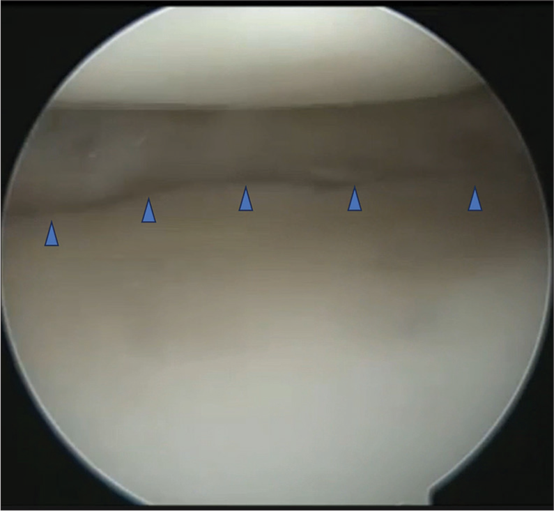
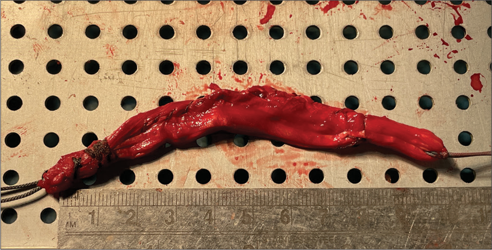
Arthroscopy with standard anterolateral and anteromedial portals showed near-complete loss of meniscus due to auto-meniscectomy with absent roots, and the patient was found suitable for meniscus transplant [Figure 1]. Loose bodies from the torn meniscal tissue were found and removed. There was an associated focal grade III cartilage injury in the medial femoral condyle. ACL graft was prepared using ipsilateral hamstring tendons. The tibial and femoral ACL sockets with tunnels were created using standard techniques. Semitendinosus graft from the opposite limb was harvested and cleaned. Using an 11-Gauge bone marrow needle (Jamshidi), 15 mL of BMA was obtained from the proximal tibia utilizing the graft harvest site without tourniquet inflation. It was placed along the edge of a dish, slowly stirred to promote the formation of a linear clot, and the serum was discarded. The round distal part of the graft was folded onto the flat proximal part along with the BMA-derived fibrin clot. The flattened part was wrapped around it and sutured like a purse with a non-absorbable 2-0 braided polyethylene suture [Figure 2]. Whip stitches using no. 5-Ethibond were placed on the two ends. Thus, a double-strand graft was prepared with BMA clot sandwiched in between. Before surgery, the length of the required graft was roughly estimated by taking measurement on the axial section of the MRI corresponding to the level of the tibial plateau on a DICOM viewer using the ”open polygon” feature. It was roughly 7 cm, and taking into account the graft portions to be inserted in the root sockets, a total graft length of around 11 cm was obtained. The thickness of the two ends of the graft was measured using a graft sizing block. A reduction suture was placed in the middle of the graft to aid in the reduction inside the joint. The capsule surface was rasped using a meniscal rasp. The posterior root was cleared. A tibial ACL drill guide was used to drill an all-inside socket of appropriate size (7 mm in this case) at the intended location of the posterior root using a flipping reverse cutting drill bit (Flipcutter, Arthrex, Florida, United States) and a passing suture was placed. The tunnel for the anterior root was drilled outside-in using the tibial ACL jig (6 mm), and another passing suture was placed. The passing sutures were brought out together from the anteromedial portal, and the posterior root, followed by the anterior root, was pulled inside their respective sockets till the previously placed mark at 20 mm, thus bringing the meniscal graft into the joint. The reduction sutures were retrieved first and pulled outward, and the meniscus was reduced toward the capsule. Multiple inside-out stitches were placed at roughly 4–5 mm intervals in vertical and oblique configurations using non-absorbable 2-0 braided polyethylene sutures to fix the posterior horn and body of the meniscus to the capsule. The posterior-most part of the posterior horn was stabilized using an all-inside repair device to avoid neurovascular injury. The anterior horn was stabilized with multiple outside-in futures using a Prolene 2-0 suture to railroad the braided polyethylene sutures. In total, 12 sutures were required: 8 inside-out sutures (superior and inferior, vertical and oblique), three outside-in sutures, and one all-inside suture. Following this, the ACL graft was passed and fixed using an adjustable loop device on the femoral side and an interference screw on the tibial side using standard technique. All the meniscocapsular fixation sutures were then tied on the capsule, utilizing a small posteromedial incision and additional stab incisions anteriorly as needed. Finally, the Ethibond sutures from the posterior and the anterior roots were tied on titanium cortical buttons. During this process, the meniscus was visualized arthroscopically to ensure that the capsule was adequately pulled centrally toward the margins of the tibial condyle. Wounds were closed after a thorough wash, and a compression bandage and a long knee splint were applied [Video 1].

- Near-total absence of meniscus tissue and absent roots due to auto-meniscectomy noted during arthroscopy (blue arrow heads).

- Final prepared graft from autologous semitendinosus tendon with bone marrow aspirate-derived fibrin clot sandwiched between the two stitched layers of graft.
Video 1:
Video 1:Video of meniscus transplant with autologous semitendinosus graft with bone marrow aspirate-derived fibrin clot along with anterior cruciate ligament reconstruction.The post-operative protocol was similar to that for a meniscal repair, with the range of motion restricted to 30°, 60°, and 90° of flexion for the first 2, 4, and 6 weeks, respectively. The patient was mobilized from day 1 but was kept non-weight-bearing for 6 weeks. After this, progressive weight-bearing and open-chain strengthening was initiated. Sports activities, twisting movements, and squatting were restricted for 6 months.
Follow-up
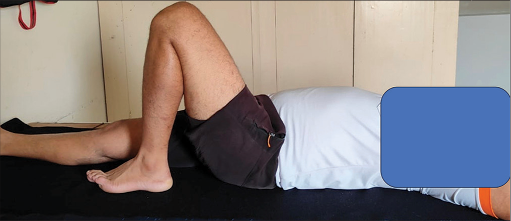
The patient obtained 0–150° range of motion by 4 months. At 6 months, he had returned to his active duties, and sports-specific training was started [Figure 3]. Lysholm’s score was 17, 76, and 88 out of 100 at pre-operative, 6 months and 9 months post-operative time periods, respectively. Visual analog scale score was 3/10 at the latest follow-up of 9 months, as compared to 8/10 reported preoperatively. MRI was done at 6 months to evaluate the condition of the graft. The signal intensities were slightly less hypointense than a normal meniscus but the meniscus graft showed reasonable uptake with characteristic triangular or wedge shape and good uptake of roots [Figure 4].

- Clinical photograph of knee showing range of motion at 9 months follow-up.

- Selected sections of magnetic resonance imaging done at 6 months follow-up. (a) Sagittal section through the middle of the medial tibial plateau showing the wedge-shaped graft with hypointense signals (blue arrows). (b) Sagittal section passing through the roots of the medial meniscus graft (blue arrows). The edge of the anterior cruciate ligament graft in the tibial tunnel is also visualized (blue arrow heads). (c) Coronal section passing through the posterior horn (blue arrow heads) and posterior root of the meniscus graft (blue arrow).
DISCUSSION
In this case report, we have presented the technique as well as early follow-up of a patient with ACL tear along with near-complete loss of medial meniscus due to auto-meniscectomy with focal grade III cartilage lesion in medial femoral condyle, managed with ACL reconstruction along with meniscus transplant with autologous semitendinosus graft with clotted BMA, in the primary sitting.
In general, meniscal transplants have been used in patients who have developed post-meniscectomy symptoms.[4] In our case, the patient presented 1-year-after the injury with focal grade III cartilage lesion and was symptomatic, with knee pain on walking in addition to complaints of instability. He had near-complete loss of meniscus and absent roots due to auto-meniscectomy, comparable to a total meniscectomy, and was considered an appropriate candidate for meniscus transplant in the first surgery itself.
It is a known fact that subtotal or total meniscectomy accelerates the development of OA.[5] Hence, we suggest that the indications of meniscus transplant should include even those patients who are undergoing a sub-total meniscectomy, have normal-looking cartilage, and have not yet developed post-meniscectomy symptoms, without waiting for them to develop post-meniscectomy symptoms or OA.
The literature on use of autologous tendon graft, particularly hamstring graft, is limited. During 1990’s, a few studies described the use of quadriceps tendon for open meniscal replacement.[6,7] However, initial reports were not encouraging and quadriceps tendon was not considered a suitable option.[8,9] In 2013, a case report described the use of semitendinosus tendon autograft for reconstructing only the meniscal wall to support a collagen meniscal implant.[10] The patient noted significant improvement in functional outcome and MRI revealed a successful integration of the implant with the capsule hence indicating the biological properties of the tendon to remodel and revascularize in the intra-articular environment.
Then, in 2017, a study investigated the use of semitendinosus tendon as autograft for meniscus reconstruction in a rabbit model and the results were promising.[11] A pilot study in 2019 reported a case of a 57-year-old female with irreparably torn discoid lateral meniscus with chondral defects, managed with meniscectomy and cartilage harvest in stage-I and autologous chondrocyte implantation and open meniscus replacement with autologous hamstring in stage-II surgery.[9] There were improvements in functional scores and a second-look arthroscopy at 1 year showed integration of meniscus graft. Rönnblad et al. used this procedure in seven symptomatic patients with previous history of meniscectomy and normal alignment to alleviate post-meniscectomy symptoms and noted improvements in IKDC score, KOOS pain subscale, and Lysholm score.[2] Similar technique was also described with peroneus graft in two patients with positive outcomes at 6–10 months.[12] In 2023, Iida et al. reported a single case of a 17-year-old woman with history of subtotal lateral meniscectomy who underwent arthroscopic meniscal transplant with autologous semitendinosus with BMA-derived fibrin clot, with good clinical and radiographic outcomes at the 24-month follow-up.[3]
The BMA-derived fibrin clot provides mesenchymal stem cells and growth factors, which have been hypothesized to promote the healing and the potential transformation into a meniscus-like tissue.[3,13] The BMA is one of the most common sources of mesenchymal stem cells for the treatment of musculoskeletal injuries.[13] In addition, this clot provides a scaffold for the healing process as well as a mechanical structure to increase the graft thickness.[3]
Thus, limited studies have reported the use of autologous hamstring meniscus transplant for compensation of absence of meniscus, with brief follow-ups but promising results.[2,3,9] The follow-up period in our study is also short; however, the radiological and clinical results in this case as well as in previous reports are promising and we strongly suggest further assessment of this procedure with longer follow-up, both in patients with post-meniscectomy symptoms as well as those young patients who are undergoing a subtotal meniscectomy in the same sitting, with the aim of preventing development of post-meniscectomy symptoms and OA.
CONCLUSION
Meniscus transplant with autologous semitendinosus tendon, along with sandwiched BMA-derived fibrin clot, was found to be an effective solution for the case with near-total loss of meniscus after 1 year of injury.
Author contributions:
HG: Concepts, design, definition of intellectual content, literature search, clinical studies, data acquisition, data analysis, manuscript preparation, and manuscript editing and review; JAJ: Manuscript preparation, data analysis, data acquisition, and literature search; JD: Literature search, clinical studies, data acquisition, data analysis, and manuscript preparation; PM: Clinical studies, data acquisition, data analysis, manuscript editing and review, and literature search.
Ethical approval
The Institutional Review Board approval is not required.
Declaration of patient consent
The authors certify that they have obtained all appropriate patient consent.
Conflicts of interest
There are no conflicts of interest.
Use of artificial intelligence (AI)-assisted technology for manuscript preparation
The authors confirm that there was no use of artificial intelligence (AI)-assisted technology for assisting in the writing or editing of the manuscript and no images were manipulated using AI.
Financial support and sponsorship
Nil.
References
- International Meniscus Reconstruction Experts Forum (IMREF) 2015 consensus statement on the practice of meniscal allograft transplantation. Am J Sports Med. 2017;45:1195-205.
- [CrossRef] [Google Scholar]
- Autologous semitendinosus tendon graft could function as a meniscal transplant. Knee Surg Sports Traumatol Arthrosc. 2022;30:1520-6.
- [CrossRef] [Google Scholar]
- Lateral meniscus autograft transplantation using hamstring tendon with a sandwiched bone marrow-derived fibrin clot: A case report. Int J Surg Case Rep. 2023;108:108444.
- [CrossRef] [Google Scholar]
- Meniscal allograft transplantation: A systematic review. Am J Sports Med. 2015;43:998-1007.
- [CrossRef] [Google Scholar]
- Does arthroscopic partial meniscectomy result in knee osteoarthritis? A systematic review with a minimum of 8 years' follow-up. Arthrosc J Arthrosc Relat Surg. 2011;27:419-24.
- [CrossRef] [Google Scholar]
- Autograft meniscus replacement: Experimental and clinical results. Knee Surg Sports Traumatol Arthrosc. 1993;1:123-5.
- [CrossRef] [Google Scholar]
- Arthroscopic and open surgical techniques for meniscus replacement--meniscal allograft transplantation and tendon autograft transplantation. Scand J Med Sci Sports. 1999;9:168-76.
- [CrossRef] [Google Scholar]
- Autogenous tendon graft substitution for absent knee joint meniscus: A pilot study. Arthrosc J Arthrosc Relat Surg. 2000;16:191-6.
- [CrossRef] [Google Scholar]
- Combined autologous chondrocyte implantation and meniscus reconstruction for large chondral defect in the lateral compartment due to discoid lateral meniscus tear: A case report. Regen Ther. 2019;10:64-8.
- [CrossRef] [Google Scholar]
- A case report of semitendinosus tendon autograft for reconstruction of the meniscal wall supporting a collagen implant. BMC Sports Sci Med Rehabil2013;. ;5:4.
- [CrossRef] [Google Scholar]
- The potential of using semitendinosus tendon as autograft in rabbit meniscus reconstruction. Sci Rep. 2017;7:7033.
- [CrossRef] [Google Scholar]
- Lateral meniscus replacement using peroneus longus tendon autograft. Arthrosc Tech. 2020;9:e1163-9.
- [CrossRef] [Google Scholar]
- Mesenchymal stem cells for tendon healing: What is on the horizon? J Tissue Eng Regen Med. 2017;11:3202-19.
- [CrossRef] [Google Scholar]


