
Translate this page into:
Dynamic MPFL reconstruction using a combination of adductor sling technique and basket weave method – A retrospective cohort study eliciting the functional results in an Indian population
*Corresponding author: Rishabh Kedia, Department of Orthopedic Surgery, Apollo Multispeciality Hospital, Kolkata, West Bengal, India. dr.rishabhkedia@gmail.com
-
Received: ,
Accepted: ,
How to cite this article: Kedia R, Basumallick MN, Khan I. Dynamic MPFL reconstruction using a combination of adductor sling technique and basket weave method – A retrospective cohort study eliciting the functional results in an Indian population. J Arthrosc Surg Sports Med. 2023;4:35-9. doi: 10.25259/JASSM_19_2023
Abstract
Objectives:
The medial patellofemoral ligament (MPFL) is one of the primary stabilizers of the patella. It resists lateral Mal-tracking of the patella and keeps the patella centered within the patellofemoral groove. MPFL reconstruction is a common and widely used procedure to treat lateral patellar instability. Most conventional techniques of MPFL reconstruction use bony tunnels in the medial femoral condyle and patella to create a static construct, which has its own set of issues. This article is aimed at describing early results from our technique for bone-sparing implant-less MPFL reconstruction with a dynamic construct using semitendinosus graft.
Materials and Methods:
This was a retrospective and single-center study of ten symptomatic patients between 12 and 35 years of age with a primary event to surgery gap of a mean of 49.6 months with a history of pain and patellar instability diagnosed by clinical and radiological means between January 2020 and August 2022. MPFL reconstruction was performed using semitendinosus graft passing under the adductor longus tendon close to its insertion and fixed at the patella with Vicryl No. 2 sutures through proximal and distal tails, respectively.
Results:
The pre-operative and follow-up mean Kujula scores were 57.5 ± 5.91 and 87 ± 4.06 (P < 0.0001), respectively, which showed significant improvement. All patients gained adequate patellar stability. No incidence of patella fracture was noted. There were no post-operative complications related to the procedure.
Conclusion:
Our study shows promising results and tries to augment the medial restraint by making the MPFL dynamic in nature, which tightens in flexion and relaxes in extension through the adductor sling at the femoral end and the basket weave technique at the patellar end. It avoids implant and bony tunnel-related complications and is simple and cost-effective.
Keywords
Medial patellofemoral ligament (MPFL)
Patellar instability
Adductor sling
Basket-weave technique

INTRODUCTION
The medial patellofemoral ligament (MPFL) is one of the primary static stabilizers of the patella.[1] The primary role of the MPFL is to resist lateral migration of the patella and to keep the patella centered within the patellofemoral groove. The MPFL is the primary ligamentous restraint of the patella, providing 50–60% of the restraining force against lateral displacement.[2]
Up to 94–100% of patients suffer from MPFL rupture after 1st-time patellar dislocation.[3]
Recurrent patellar instability can be caused by multiple anatomical factors, the most important of which is MPFL incompetence. Surgery is required both for 1st-time dislocation with osteochondral fractures[4] and recurrent instability with MPFL incompetence. Multiple tests (apprehension test, sulcus sign, J sign, etc.) have been described to check clinically for MPFL laxity.
MPFL reconstruction is a common and widely used procedure to treat lateral patellar instability, including recurrent lateral dislocation of the patella. Many different surgical techniques have been reported for MPFL reconstruction, but opinions regarding graft choice, type of fixation, graft positioning, and correct tension remain variable.[5] Because relatively young patients are most commonly affected by recurrent patellar instability after lateral patellar dislocation, the long-term clinical outcome of any surgical procedure is important.
Most conventional techniques of MPFL reconstruction use bony tunnels in the medial femoral condyle and patella to create a static construct. The common disadvantages of a static construct are over-tightening, inadequate tightening, and progressive stretching of graft.[6] There were issues with bony tunnels as well, such as widening, damage to the growth plate, and patellar fracture. As earlier methods are static, they were associated with an increased incidence of knee stiffness.[7] In our study, we have used the femoral end graft fixation through the adductor magnus tendon close to its insertion at the adductor tubercle to create a dynamic fixation point. The patellar end graft fixation is done through the quadriceps tendon near its insertion at the proximal part of its insertion and subperiosteally at the level of the equator of the patella.[7] The reconstructed MPFL after that progressively tightens in the first 20–30° flexion of the knee. As this is an implantless technique, hardware-related issues such as infections, widening of tunnels, and loosening of implants are avoided.
This article is aimed at describing short-term results from our technique for bone-sparing implantless MPFL reconstruction with semitendinosus graft (adductor sling technique, double-stranded reconstruction, and soft-tissue fixation in the extensor apparatus).
Various other factors are involved in the patellar instability, which includes trochlear dysplasia (85%, 96%), increased lateral patellar tilt (83%), patella alta (24%), and an increase in the distance between tibial tuberosity and center of the trochlear groove (TT-TG) in 56% of cases, as well as other less frequent problems/increased Q angle.[8,9] These factors have been excluded from our study as these are alignment-based bony issues, and therefore, the line of management is different from soft-tissue laxity or rupture of MPFL.
MATERIALS AND METHODS
This was a retrospective and single-center study of ten symptomatic patients between 12 and 35 years of age with a primary event-to-surgery gap of 49.6 months with a history of pain and patellar instability diagnosed by clinical and radiological means (using radiographs, scanogram, and magnetic resonance imaging) between January 2020 and August 2022.
Primary outcome measures were recurrence of patellar subluxation, dislocation, or apprehension. The secondary outcome measures included the Kujala score and Tegner score at the final follow-up.[10-12]
We calculated categorical variables that were presented as frequencies and percentages. Continuous variables were presented as mean values and standard deviation (SD) or range. A paired t-test was used to compare the Kujala and Tegner scores preoperatively and at the final follow-up. Exclusion criteria were as follows: A Q angle more than 20°; trochlear dysplasia (Dejour grade B, C, and D) or patella alta (Caton-Deschamps index of >1.3); TT-TG distance more than 20 mm; and multi ligamentous knee injury; previous operation on the same knee or any other knee pathology/atraumatic patellofemoral instability.
Surgical technique
Arthroscopy
A diagnostic arthroscopy of the knee was performed routinely to look for any associated meniscal or ligamentous injury. The articular surface is examined for any chondral lesion. If the MPFL injury is associated with an osteochondral lesion of the patella, the fragment is either fixed or excised.[4]
Harvest of semitendinosus graft
Patients were examined under anesthesia and by arthroscopy to confirm patellar instability. Harvest of the semitendinosus graft was done by standard technique through an oblique incision 4–5 cm below the medial tibiofemoral joint line. The muscle tissues are removed from the tendon graft, and the graft is prepared by tying non-absorbable sutures at both ends.
MPFL reconstruction
A 2 cm long incision is made over the medial epicondyle of the femur and deepened to the capsule [Figure 1]. The adductor tendon insertion at the adductor tubercle of the medial femoral condyle is identified by blunt dissection. The semitendinosus graft is passed under the adductor magnus tendon close to its insertion. This forms the dynamic sling fixation for the new MPFL [Figure 2].

- Skin marking over the medial femoral epicondyle depicting a 2 cm long incision site.
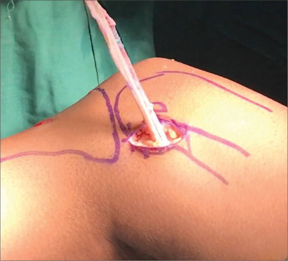
- Semitendinosus graft being passed under the adductor longus tendon close to its insertion site.
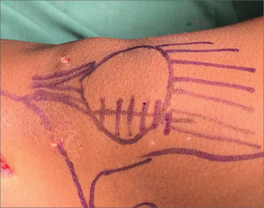
A second longitudinal incision (3 cm) is made over the patella in the midline of the knee [Figure 3]. This is deepened further to incise the deep fascia over the knee, staying superficial to the capsule. Dissecting deep to the deep fascia, a tunnel is created between layers II and III up to the first incision on the medial aspect of the knee. Through this tunnel, the free ends of the loop semitendinosus graft are recovered to the anterior incision. The proximal tail of the graft is woven through the quadriceps tendon near its insertion into the patella, creating a tunnel at the proximal pole of the patella [Figure 4]. The graft is sutured to the quadriceps tendon at the passage points with Vicryl number 2. Subperiosteal tunnels are created over the equator of the patella with a sharp Awl. The second tail of the graft is woven through this tunnel [Figure 5]. At this point, the tension is adjusted to allow for the central position and tilt of the patella with appropriate medial-lateral glide. The fixation is done at 0°, allowing for one quadrant lateral translation and the final position and tilt are checked under arthroscopic vision during the initial 30° of flexion.
- Skin marking over the patella in the midline of the knee depicting the site of the second incision.

- The proximal tail of the graft being woven through the quadriceps tendon near its insertion to the patella, creating tunnels at the proximal pole of the patella.

- The second tail of the graft being woven at the level of the equator of the patella subperiosteally.
The distal tail of the graft is then sutured to the periosteal tissue of the patella, and any excess graft is excised. Finally, the sling at the adductor tendon loop is sutured with absorbable sutures. The three incisions are finally closed, and a heavy compression dress is applied.
Post-operative rehabilitation
The patients are immediately mobilized with protected weight-bearing with a rigid knee brace and with the help of crutches. Range of motion exercises are started immediately with 0–30° of flexion for the first three weeks, increasing to 90° at 3–6 weeks subsequently, and the goal is to achieve full range of motion after that. Patients are allowed to return to sports at six months.
RESULTS
A total of ten patients (two males and eight females) were evaluated. The mean age of patients at the time of injury was 23.60 years (12–35, SD 7.50), with mean follow-up which was 51.10 months (9-120, SD 35.54). Three patients with osteochondral fractures of the patella were treated with excision as the fragments were too small to fix. The pre-operative mean Kujula score was 57.5 ± 5.91. The demographic data are shown in Table 1. The mean Kujula score at follow-up was 87 ± 4.06 (P < 0.0001), which showed significant improvement as compared to its pre-operative score. The pre-operative and follow-up Tegner scales were 3.10 and 3.40, respectively (P = 0.083), which did not show significant improvement. There were no other major post-operative complications related to the procedure or rehabilitation. There was no recurrence of instability of the patella over the follow-up period. Significant and sustained improvement in pain was observed over follow-up periods. Post-operative complications were rare, with one patient having terminal restriction in flexion.
| Total patients | 10 |
| Male | 2 |
| Female | 8 |
| Side | |
| Right | 6 |
| Left | 4 |
| Type of dislocation | |
| Traumatic | 6 |
| Recurrent | 4 |
| Mean age at surgery (years) | 23.6 (12-35) |
| Mean follow-up (months) | 51.1 (12-120) |
| Duration between 1st episode and intervention (months) | 49.6 (1-120) |
| Pre-operative mean Kujala’s score | 65.5±17.0 |
DISCUSSION
Recurrent instability after patellar dislocation remains the main indication for surgical reconstruction of MPFL. Multiple methods have been described for the reconstruction of the medial constraints of the patella, from medial plication to ligament reconstruction. MPFL reconstruction remains the mainstay of the treatment. The hamstring (gracilis or semitendinosus) remains the most commonly used autograft for MPFL reconstruction. Other grafts, such as quadriceps tendon, adductor magnus graft, and patellar tendon graft, have also been used.[13,14] There are multiple techniques described for the fixation of grafts which may be broadly divided into fixation by implants such as anchor sutures or interference screws and implant-less fixation with the help of sutures through bone tunnels. Complications of MPFL reconstruction techniques include recurrent dislocation, subluxation, patellar fracture, improper placement of the graft, positive apprehension test, and over-tightening, leading to stiffness and pain.[15,16]
Functional evaluation was done using the Kujala scoring system and the Tegner activity scale in the present study. The pre-operative and follow-up mean Kujula score was 57.5 ± 5.91 and 87 ± 4.06, respectively, which showed significant improvement (P < 0.0001). The previous studies using implants revealed similar results, with the mean postoperative Kujala score being 84 in Christiansen et al. study,[17] and 90.9 in Song et al.[18] In a study done by Shimizu et al. using implantless techniques, the Kujula score was 86.7 which was comparable with our results.[5] Schneider et al. reported a mean post-operative Kujala score of 85.8.[19]
The pre-operative and follow-up Tegner scores were similar as most of our patients were not highly demanding in terms of activity, so the intervention relieved them of their symptoms but did not improve their level of activity.
In our study, there were no dislocations, subluxations, or positive apprehension reported. Two cases were reported to have initial terminal restriction of flexion, which improved over subsequent months. The advantage of this technique was the lack of complications due to hardware, such as impingement, bone resorption and tunnel widening, infection, and persistent knee pain.
This method was aimed at restoring the dynamics of the MPFL. As compared to previous studies by Schöttle et al.[20] which were focused on radiological landmarks; this study focused on recreating the dynamics, thus having an advantage of minimizing the radiological error and benefiting skeletally immature children, as Schöttle point was found to be incorrect.[7]
As compared to previous studies by Kodkani in which the graft is looped up to its center underneath this ligamentousperiosteal sleeve,[7] in our technique, the adductor sling helps make this form of reconstruction more dynamic as the adductor tendon would help the reconstructed MPFL to contract in the initial degrees of flexion making it less susceptible for dislocation in the initial degrees of flexion.
Unlike the commonly used fixation methods using implants such as screws or anchors, this soft-tissue fixation technique is more dynamic and does not result in a rigid construct.
As this procedure is entirely based on soft tissue fixation, there are no hardware and bony tunnel-related complications such as anterior knee pain, hardware-related allergies, or infections. As implants make the construct more rigid, tightening can lead to stiffness and restriction in the range of motion of the knee.
In this technique, the insertion is double-stranded, and both strands are tensioned at variable degrees of flexion; this provides stability at variable degrees of flexion as compared to other methods having one strand of insertion.
CONCLUSION
This study shows promising results and tries to augment the medial restraint by making the MPFL dynamic in nature, which tightens in flexion and relaxes in extension through the adductor sling at the femoral end and the basket weave technique at the patellar end. It is a simple and cost-effective technique and avoids implant and bony tunnel-related complications such as loosening, allergies/infection, anterior knee pain, or cut-through of the implant through bone.
Ethical approval
The Institutional Review Board approval is not required.
Declaration of patient consent
The authors certify that they have obtained all appropriate patient consent.
Conflicts of interest
There are no conflicts of interest.
Use of artificial intelligence (AI)-assisted technology for manuscript preparation
The authors confirm that there was no use of artificial intelligence (AI)-assisted technology for assisting in the writing or editing of the manuscript and no images were manipulated using AI.
Financial support and sponsorship
Nil.
References
- Implantless patellar fixation in medial patellofemoral ligament reconstruction. Chin J Traumatol. 2019;22:281-5.
- [CrossRef] [PubMed] [Google Scholar]
- First-time patellar dislocation: Surgery or conservative treatment? Sports Med Arthrosc Rev. 2012;20:128-35.
- [CrossRef] [PubMed] [Google Scholar]
- Acute patellar dislocation. What to do? Knee Surg Sports Traumatol Arthrosc. 2013;21:275-8.
- [CrossRef] [PubMed] [Google Scholar]
- Traumatic patellar dislocation: Loose bodies and the MPFL. J Pediatr Orthop. 2012;32(Suppl 1):S47-51.
- [CrossRef] [PubMed] [Google Scholar]
- Middle-to long-term outcome after medial patellofemoral ligament reconstruction with Insall's proximal realignment for patellar instability. Asia Pac J Sports Med Arthrosc Rehabil Technol. 2019;17:5-9.
- [CrossRef] [PubMed] [Google Scholar]
- Technical errors during medial patellofemoral ligament reconstruction could overload medial patellofemoral cartilage: A computational analysis. Am J Sports Med. 2006;34:1478-85.
- [CrossRef] [PubMed] [Google Scholar]
- "Basket weave technique" for medial patellofemoral ligament reconstruction: Clinical outcome of a prospective study. Indian J Orthop. 2016;50:34-42.
- [CrossRef] [PubMed] [Google Scholar]
- The role of trochlear dysplasia in patellofemoral instability. J Am Acad Orthop Surg. 2011;19:8-16.
- [CrossRef] [PubMed] [Google Scholar]
- MR imaging of patellar instability: Injury patterns and assessment of risk factors. Radiographics. 2010;30:961-81.
- [CrossRef] [PubMed] [Google Scholar]
- Reliability and validity of the anterior knee pain scale: Applications for use as an epidemiologic screener. PLoS One. 2016;11:e0159204.
- [CrossRef] [PubMed] [Google Scholar]
- The reliability, validity, and responsiveness of the Lysholm score and Tegner activity scale for anterior cruciate ligament injuries of the knee: 25 years later. Am J Sports Med. 2009;37:890-7.
- [CrossRef] [PubMed] [Google Scholar]
- Tegner level is predictive for successful return to sport 2 years after anterior cruciate ligament reconstruction. Knee Surg Sports Traumatol Arthrosc. 2021;29:3010-6.
- [CrossRef] [PubMed] [Google Scholar]
- Medial patellofemoral ligament reconstruction: Fixation strength of 5 different techniques for graft fixation at the patella. Arthroscopy. 2013;29:766-73.
- [CrossRef] [PubMed] [Google Scholar]
- MPFL reconstruction using a quadriceps tendon graft: Part 2: Operative technique and short term clinical results. Knee. 2014;21:1175-9.
- [CrossRef] [PubMed] [Google Scholar]
- A systematic review of complications and failures associated with medial patellofemoral ligament reconstruction for recurrent patellar dislocation. Am J Sports Med. 2012;40:1916-23.
- [CrossRef] [PubMed] [Google Scholar]
- Technical failure of medial patellofemoral ligament reconstruction. Arthroscopy. 2011;27:1153-9.
- [CrossRef] [PubMed] [Google Scholar]
- Reconstruction of the medial patellofemoral ligament with gracilis tendon autograft in transverse patellar drill holes. Arthroscopy. 2008;24:82-7.
- [CrossRef] [PubMed] [Google Scholar]
- Anatomic medial patellofemoral ligament reconstruction using patellar suture anchor fixation for recurrent patellar instability. Knee Surg Sports Traumatol Arthrosc. 2014;22:2431-7.
- [CrossRef] [PubMed] [Google Scholar]
- Outcomes after isolated medial patellofemoral ligament reconstruction for the treatment of recurrent lateral patellar dislocations: A systematic review and meta-analysis. Am J Sports Med. 2016;44:2993-3005.
- [CrossRef] [PubMed] [Google Scholar]
- Radiographic landmarks for femoral tunnel placement in medial patellofemoral ligament reconstruction. Am J Sports Med. 2007;35:801-4.
- [CrossRef] [PubMed] [Google Scholar]


