Translate this page into:
Migration of patella tension band metal wires to popliteal fossa: Retrieval by posterior knee arthroscopy – A case report and literature review
*Corresponding author: Narendran Pushpasekaran, Department of Orthopaedics, Vinayaka Mission’s Kirupananda Variyar Medical College and Hospital, Vinayaka Mission’s Research Foundation (Deemed to be University), Salem, Tamil Nadu, India. drnaren247ortho@gmail.com
-
Received: ,
Accepted: ,
How to cite this article: Pushpasekaran N, Karthik CA, Muthukannan HS. Migration of patella tension band metal wires to popliteal fossa: Retrieval by posterior knee arthroscopy – A case report and literature review. J Arthrosc Surg Sports Med. 2023;4:53-5. doi: 10.25259/JASSM_15_2023
Abstract
Breakage of metal wires after fixation of patella fractures are common. However, migration of the broken wires to intraarticular location, popliteal fossa and distant sites can be least expected and a difficult scenario for retrieval. We describe in a 58-year male who presented with recurrent locking and migration of broken wires in popliteal fossa. he had fixation of patella fracture nine years ago. The metal wire was retrieved safely from the posterior lateral aspect of knee popliteal fossa by posterior knee arthroscopy using two posterior lateral portals. Through this case, we review the presentation of migrated wires, methods of retrieval and role of arthroscopy. Retrieval of migrated wires by knee arthroscopy can be successful in intraarticular location and in select cases at popliteal fossa, however with significant challenges. To avoid concerns with migration of wires, patients treated with wires for patella fractures should be adequately followed with radiography and sensitized for removal before breakage or at the time of breakage.
Keywords
Patella fractures
Tension band wires
Wire breakages
Popliteal fossa
Knee arthroscopy
Migration of wires
Posterior knee arthroscopy
INTRODUCTION
Tension band wires (TBWs) have been the gold standard for the management of patella fractures. Cerclage wires are added to prevent the comminuted fragments from displacements till union. Although the breakage of wires is common, migration of the components to intra-articular, popliteal fossa, and distant sites is least expected and occasionally reported in the literature.[1-3] Removal of the same from the popliteal fossa is cumbersome and requires different open approaches. We describe a case where the migrated metal wire in the posterior lateral aspect of the knee popliteal fossa caused impingement on squatting and was removed successfully by posterior knee arthroscopy, unlike the previously described open methods in popliteal fossa.
CASE REPORT
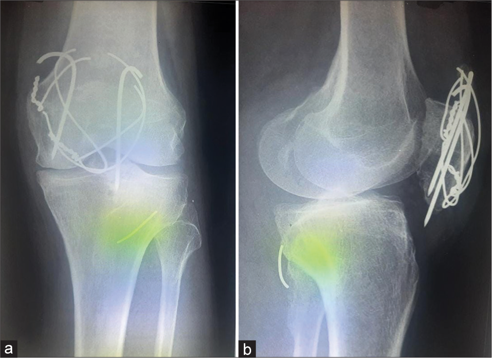
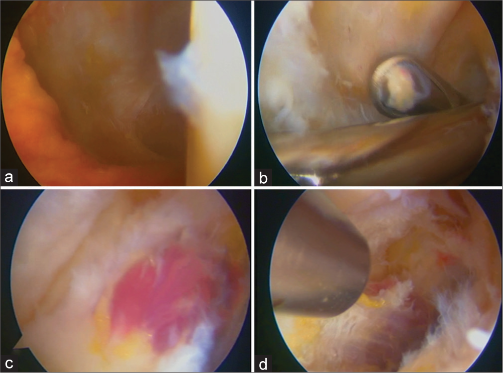
A 58-year-old laborer had occasional anterior knee pain since the open reduction and internal fixation of a comminuted patella fracture nine years ago. Although his symptoms were relieved by medications and rest, he noticed disturbances in the posterior aspect of his knee while squatting for about four months. Clinical examination of the knee revealed a previous linear scar in the anterior aspect, tenderness in the popliteal fossa, patellofemoral joint crepitus, and anterior knee swelling. Plain radiographs of the knee showed a united patella fracture with Kirschner wires (K-wires), broken TBW, and cerclage wires [Figure 1]. Another metal piece was observed in the posterior lateral aspect of the knee and corresponded to the gap of TBW. The implants around the patella were removed through the anterior approach. Diagnostic arthroscopy was performed through anterior lateral and anterior medial portals. The posterior lateral compartment was viewed with the scope between the anterior cruciate ligament and the lateral femoral condyle [Figure 2]. A 5 mm rent was observed in the posterior capsule. Two posterior lateral portals separated by about 1.5 cm were established for further exploration in the posterior aspect of the knee. The metal wire was found within the bulk of the popliteal muscle, and by gentle manipulation with an arthroscopic grabber, the tip of the metal wire was visualized, held, and removed successfully [Figure 3] and [Video 1]. There were no peroneal nerve or arterial injuries observed in the post-operative period. He had returned to his squatting habits and was relieved of his previous symptoms.

- Broken and migrated wires of the tension band wire construct. (a) Anterior, posterior view showing missing wire at the superior border of the patella. (b) Lateral view showing the migrated wire in the posterior lateral aspect of the knee.

- Diagnostic knee arthroscopy and portals. (a) The posterior lateral capsule showing rent. (b) Two posterior lateral portals were made for posterior knee arthroscopy. (c and d) View from the posterior lateral portal showing absent wires from the surface of the popliteus tendon.

- Arthroscopic posterior knee exploration and retrieval. (a) The metal wires are seen within the bulk of the popliteal muscle and the synovial layer around it. (b) Attempted removal revealed multiple fibrous bands entrapping the wire. (c) The tip of the wire is visualized and removed. (d) Post-operative radiographs showing complete removal of metal wires.
Video 1:
Video 1:Migrated metal hardware from patella to popliteal fossa. Exploration and retrieval of the hardware by posterior knee arthroscopy.DISCUSSION
Breakage of metal wires used for fixation and reconstruction of patella fractures is not uncommon; however, migration of the wires to sites distant from patella is are least expected and a difficult situation due to the requirement of additional incisions and extensive surgery for removal.[1] The metal wires have been reported to migrate through the intra-articular route or the subretinacular layer to the posterior aspect of the knee and have been retrieved from intra-articular locations, popliteal fossa, and ankle.[1-3] In this case, the broken wire was retrieved from the posterior lateral aspect of the knee within the bulk of the popliteal muscle.
Active individuals, improper tensioning principles and deep location of the wires in the quadriceps tendon have been a common concern with the breakage and migration of metal wires. Breakage of K-wires, cerclage wires, and the figure of eight wires have all been reported.[4] The patient described in this case was a muscular individual who had a fixation with two K-wires, cerclage wires, and a figure of eight wires for a comminuted fracture of the patella. Inadequate tensioning principles can also be considered in this case, as the wires had spirals on only one side. The cerclage wire and TBW had broken segmentally, but the broken fragment of TBW at the superior pole of the patella had migrated to the posterior aspect of the knee, probably through the intra-articular route, as rent was observed in the posterior capsule.
Longer intervals from the initial fixation of patella fractures have been an independent factor for the breakage of wires. Sammy, in a retrospective analysis of 59 patients who had removed patella TBW construct for various reasons, observed that 31 patients (53%) had wires broken at the time of removal, including seven patients with migrated wires. The mean duration at the time of removal of broken wires was 74 months from the initial fixation,as against the 28 patients who had no breakage of wires at the time of removal in 19.4 months.[5] This correlated with our patient, who had presented nine years after the initial fixation and had broken wires at two sites and migration into the popliteal fossa. Removal of TBW implants posts union of patella fracture by two years from initial fixation and regular follow-up with radiographs to look out for wire breakages and appropriate removal on noticing the broken wires without further delay can prevent migration of wires.
Choi et al. retrieved migrated wires from two patients in a similar location, as in our case, at the posterior lateral aspect of the knee popliteal fossa. In one case, they had attempted to remove the fragment arthroscopically but failed and had to do a direct lateral approach to the knee to retrieve the migrated wire. In another case, they had done an extensive posterior approach to the knee to retrieve the migrated fragments.[6] Similarly, Meena et al. reported K-wire migration into the popliteal fossa that was retrieved by open method (lateral approach to knee).[7] We had done meticulous posterior knee exploration by arthroscopy through two posterior lateral portals and safely retrieved the wires from the muscle of the popliteus tendon. We did not observe any neurovascular deficit.
CONCLUSION
Patella fractures treated with TBWs and cerclage wires require radiographs at regular intervals to determine union and wire breakages. Broken wires could be removed at the earliest to avoid migration. Migrated wires to the intraarticular and posterior knee can be retrieved arthroscopically with minimal risks.
Ethical approval
The Institutional Review Board approval is not required.
Declaration of patient consent
Patient’s consent not required as patient identity is not disclosed or compromised.
Conflicts of interest
There are no conflicts of interest.
Use of artificial intelligence (AI)-assisted technology for manuscript preparation
The authors confirm that there was no use of artificial intelligence (AI)-assisted technology for assisting in the writing or editing of the manuscript and no images were manipulated using AI.
Video available on:
Financial support and sponsorship
Nil.
References
- Arthroscopic removal of a wire fragment from the posterior septum of the knee following tension band wiring of a patellar fracture. Case Rep Orthop. 2015;2015:827140.
- [CrossRef] [PubMed] [Google Scholar]
- Migration of a broken cerclage wire from the patella into the heart. A case report. J Bone Joint Surg Am. 2006;88:2057-9.
- [CrossRef] [PubMed] [Google Scholar]
- Migration of a Kirschner wire to the dorsolateral side of the foot following osteosynthesis of a patella fracture with tension band wiring: A case report. J Med Case Reports. 2016;10:41.
- [CrossRef] [PubMed] [Google Scholar]
- Intra-articular migration of broken patellar tension band wire: A rare case. J Orthop Case Rep. 2016;6:41-3.
- [Google Scholar]
- Breakage and migration of metal wires in operated patella fractures: Does it correlate with time? J Orthop Trauma Rehabil. 2013;17:13-6.
- [CrossRef] [Google Scholar]
- Migration to the popliteal fossa of broken wires from a fixed patellar fracture. Knee. 2008;15:491-3.
- [CrossRef] [PubMed] [Google Scholar]
- Delayed migration of K-wire into popliteal fossa used for tension band wiring of patellar fracture. Chin J Traumatol. 2013;16:186-8.
- [Google Scholar]


