
Translate this page into:
Suture disc adjustable suspensory fixation device – A cost-effective technique for tibial cortical fixation in cruciate ligament reconstruction
*Corresponding author: Sanjay Singh, Expert Ortho Clinic, Mumbai, Maharashtra, India. singsp@gmail.com
-
Received: ,
Accepted: ,
How to cite this article: Singh S. Suture disc adjustable suspensory fixation device – A cost-effective technique for tibial cortical fixation in cruciate ligament reconstruction. J Arthrosc Surg Sports Med. 2023;4:60-3. doi: 10.25259/JASSM_21_2023
Abstract
Cruciate ligament reconstruction with adjustable suspensory devices at the femur and tibia allows graft tensioning at both ends, but it is costly due to the tibial suspensory device. To overcome the cost, the author describes a technique where the adjustable suspensory cortical fixation device can be made with a suture disc and an ultra-high-molecular-weight polyethylene suture, which is simple, cost-effective, and making it reproducible even with less experienced arthroscopy surgeons.
Keywords
Suture disc adjustable loop
Tibial suspensory fixation
Cost-effective double suspensory fixation

INTRODUCTION
Suspensory cortical fixation devices on the tibial side in the anterior cruciate ligament (ACL) and posterior cruciate ligament reconstruction have emerged as a method of fixation as an alternative to interference screw fixation, especially with the advent of all inside techniques.[1]
There are many advantages of using suspensory cortical fixation at both ends, as they are re-tensionable devices the final tightening of the soft-tissue graft can be readjusted from both ends until it is fully taut, there is no requirement of long length of the grafts as the fixation of graft is at the cortex instead of being in the bone and this gives an opportunity to make the graft thicker by increasing the folds of the tendons passing through both the ends of suspensory devices. Although very convenient, there is an added cost of the adjustable loop implant at the tibial end which increases the cost of the ligament reconstruction significantly. This cost difference is important in our country where less affording patients cannot bear the additional cost.
To overcome the cost issue, the author has come up with an adjustable suspensory fixation device, which is made on the operating table using a suture disc and a No. 2 ultra-high-molecular-weight polyethylene (UHMWPE) suture.
MAKING OF THE ADJUSTABLE SUSPENSORY DEVICE
The extra UHMWPE suture is removed from the femoral adjustable loop and is substituted with a No. 2 Ethibond suture (Ethicon, Bridgewater, NJ) for flipping the button.
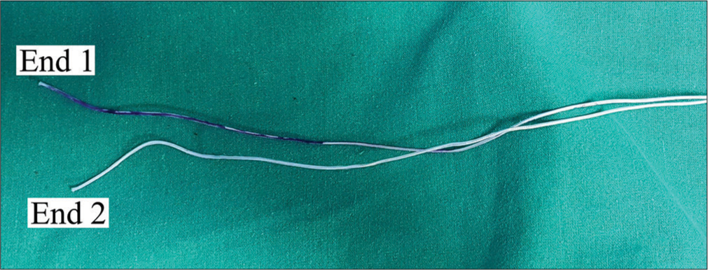
The free ends of the harvested tendon/tendons (Semitendinosus ± Gracilis, if quadrupled Semitendinosus is <8 mm in diameter) are whip-stitched together using a No. 2 UHMWPE suture [Figure 1].

- Both ends of the Semi T are Whip stitched together.
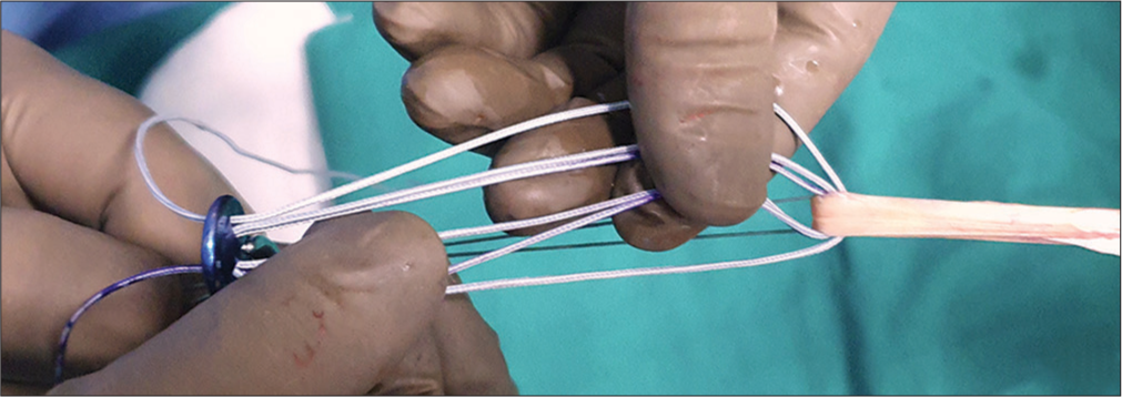
The removed UHMWPE suture is now passed first through the loop end of the tendon so that the free ends of the suture exit through the two separate holes of the suture disc. The assistant holds the whip-stitched end of the tendon on one side and the suture disc with artery forceps on the other side such that the holes are accessible to pass sequential throws of the free ends [Figure 2 and Supplementary Video 1].
- Free ends of the UHMWPE suture to be used for making the adjustable loop (one end is marked for demonstration purposes). UHMWPE: Ultra-high-molecular-weight polyethylene.
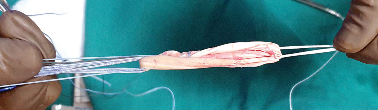
End No. 1 is now passed through the adjoining hole to go through the tendon and exit back again from the 1st hole, making two strands of UHMWPE loop connecting the tendon and the suture disc on each side. Similar steps are done with the opposite end of the suture, making three strands of UHMWPE suture on each side. This completes the formation of the adjustable loop over a suture disc [Figure 3 and Supplementary Video 1].
- 3 strands of UHMWPE suture on each side mark the completion of the adjustable loop device. UHMWPE: Ultra-high-molecular-weight polyethylene.
The whip-stitched end of the graft is now docked back towards the suture disc with the femoral adjustable loop sitting in between [Figure 4], making it a quadrupled/eightfold graft. The graft is now sutured near both ends with the suture material of the surgeon’s choice to make is a single incorporated graft, which is now put for tensioning on the graft preparation board [Figure 5].
- Docking of the whip-stitched end towards the suture disk over an adjustable femoral button to make a four or 8-strand graft.
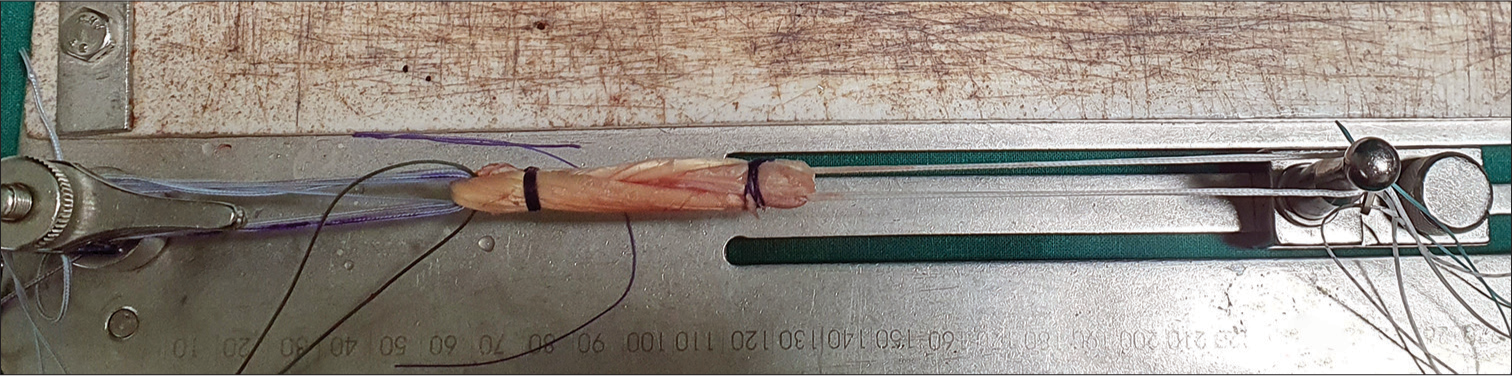
- The graft is sutured at both ends to make it a single incorporated graft and tensioned on the graft preparation board.
OPERATIVE TECHNIQUE
The femoral and tibial tunnels are drilled as usual; while passing the graft, one needs to be careful about not pulling on the free ends coming out of the suture disc as that will lead to a reduction of the loop; instead one can hold the loop just above the suture disc [Figure 6] with a hemostat loosely clamped to it. Once the graft is reduced in the femoral socket, the knee is cycled for 20–25 flexion extensions, then keeping the knee in 20–30° flexion, posterior drawer force is applied on the proximal tibia now the free ends of the prepared adjustable suture disc loop are pulled one by one to reduce the suture disc to the tibial tunnel orifice and this tightens the graft from the tibial end as well giving good tension on the graft and the suture disc is snug fit against the cortex. Alternate half hitches are tied to secure the loop [Supplementary Video 1].
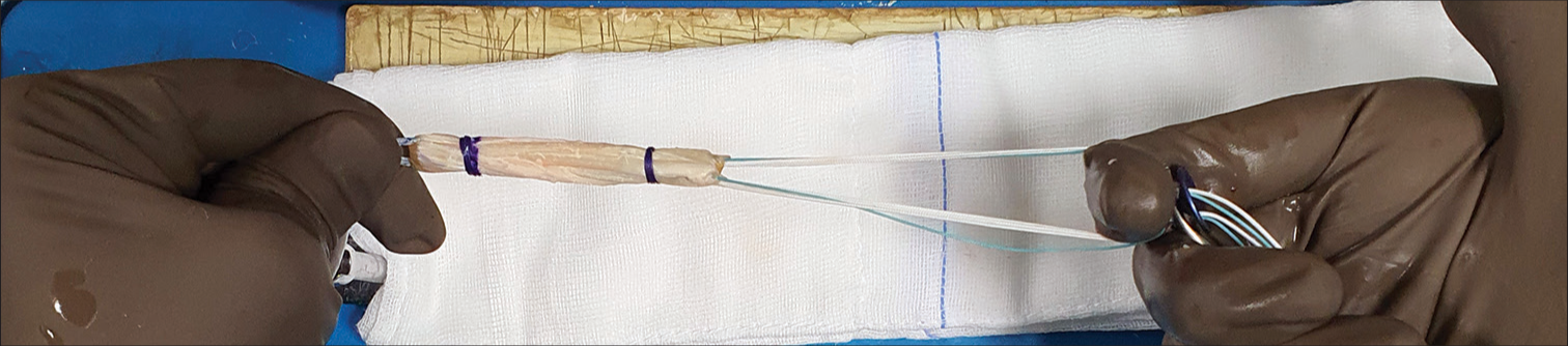
- While inserting the graft tibial end to be held with a finger between the strands on each of the suture disk holes with a lightly clamped hemostat.
The arthroscope is put in the joint to check on the tension of the graft, and if the graft needs re-tensioning, it can be done at the femoral side. Closure is done as usual.
DISCUSSION
Adjustable cortical suspensory fixation at the tibia, as compared to using an Interference screw, gives the opportunity to re-tension the graft at both ends and avoid problems of interference screws. Interference screws, bioabsorbable or metallic, have their problems of cyst formation,[2] tunnel widening.[3] All inside ACL reconstruction is expensive.[4] There are not many studies showing the use of cortical suspensory devices on the tibial side as solitary fixation with the complete tibial tunnel. Colombet et al.[5] showed better conditions for graft ligamentization within the tibial tunnel at six months in the adjustable loop group versus the absorbable interference screw group [Table 1]. Considering the study, an adjustable loop device can be used on the tibia with a complete tibial tunnel [Table 1]. This reduces the additional cost of a retro drill, and with the authors preferred technique mentioned above, the cost can further be brought down using a simple suture disk and a No. 2 or No. 5 UHMWPE suture which in combination is again cheaper than a conventional adjustable loop device; [Table 1] in addition, this technique provides a secure fixation on the tibia with a suture disc attached to 8 strands of UHMWPE sutures making it a robust construct which is re-tensionable, in case of fixed loop used at femur this technique allows retensioning from the tibial end [Table 1].
| Pearls | Pitfalls |
|---|---|
| Easy to make adjustable device on the table with available resources | Graft preparation takes more time compared to the conventional interference screw fixation technique |
| Removes slack from the tibial end of the graft in case of fixed loop at the femur | |
| Low cost | |
| Ensures circumferential fit of the tunnel by the graft | |
| It uses a conventional complete tibial tunnel, which is easily reproducible. |
CONCLUSION
This is a simple, cost-effective, and reproducible technique to make an adjustable loop cortical fixation device for tibial fixation in cruciate ligament reconstruction. Along with the opportunity of re-tensioning the graft at the tibial end, this technique provides robust tibial fixation as well.
Ethical approval
The Institutional Review Board approval is not required.
Declaration of patient consent
Patient’s consent not required as patients identity is not disclosed or compromised.
Conflicts of interest
There are no conflicts of interest.
Use of artificial intelligence (AI)-assisted technology for manuscript preparation
The authors confirm that there was no use of artificial intelligence (AI)-assisted technology for assisting in the writing or editing of the manuscript and no images were manipulated using AI.
Video available on:
Financial support and sponsorship
Nil.
References
- All-inside anterior cruciate ligament graft-link technique: Second-generation, no-incision anterior cruciate ligament reconstruction. Arthroscopy. 2011;27:717-27.
- [CrossRef] [PubMed] [Google Scholar]
- Bioabsorbable interference screw failure in anterior cruciate ligament reconstruction: A case series and review of the literature. Knee. 2015;22:256-61.
- [CrossRef] [PubMed] [Google Scholar]
- ACL reconstruction with adjustable-length loop cortical button fixation results in less tibial tunnel widening compared with interference screw fixation. Knee Surg Sports Traumatol Arthrosc. 2020;28:1036-44.
- [CrossRef] [PubMed] [Google Scholar]
- Material costs of anterior cruciate ligament reconstruction with hamstring tendons by two different techniques. Orthop Traumatol Surg Res. 2013;99:196-201.
- [CrossRef] [PubMed] [Google Scholar]
- Incorporation of hamstring grafts within the tibial tunnel after anterior cruciate ligament reconstruction: Magnetic resonance imaging of suspensory fixation versus interference screws. Am J Sports Med. 2016;44:2838-45.
- [CrossRef] [PubMed] [Google Scholar]


