
Translate this page into:
Recent advances and future trends in hip arthroscopy


*Corresponding author: Vikas Khanduja, Cambridge Hip Preservation Service, Trauma and Orthopaedics, Addenbrooke’s - Cambridge University Hospital, Box 37, Hills Road, Cambridge CB2 0QQ, United Kingdom. vk279@cam.ac.uk
-
Received: ,
Accepted: ,
How to cite this article: Zhang J, Pettit M, Sunil Kumar KH, Khanduja V. Recent advances and future trends in hip arthroscopy. J Arthrosc Surg Sport Med 2020;1(1):81-9.
Abstract
Hip arthroscopy (HA) is a well-established minimally invasive technique used to treat a wide range of conditions. Technological innovations have expanded the scope of HA and improved its outcomes. Several authors have described post-less arthroscopy to overcome the risk associated with the use of the perineal post for obtaining distraction of the hip joint. Instrument refinement has improved the efficacy of labral repair and reconstruction. Several treatment strategies are available for articular cartilage defects including ACI, MACI, and mosaicplasty, to name a few. Some authors have suggested that ligamentum teres reconstruction is helpful in young active patients with femoroacetabular impingement experiencing subluxation of the hip joint. The incorporation of new technology and patient-specific approaches represents a new way to improve HA outcomes. Virtual reality simulation training is the key to overcoming the steep learning curve associated with HA and to achieve high standards early in a surgeon’s career. Computer-based navigation surgery may be the key to accurately resecting the cam deformity and reducing the rate of revision surgery due to inaccurate resection at the index procedure. Assessment and optimization of the baseline psychological state and effective pre-habilitation may also improve outcome measures. Adequate stratification of the pathology and assessment of baseline patient characteristics influences the long-term outcome following the HA.
Keywords
Arthroscopy
Virtual reality
Innovations
Navigation
Outcome

INTRODUCTION
Hip arthroscopy (HA) is a well-established, minimally invasive surgical technique used to treat intra- and extra-articular disorders of the hip, such as femoroacetabular impingement (FAI), labral and chondral injuries, ligamentum teres injuries, iliopsoas impingement, sub- spinous impingement, and the indications continue to expand. Compared with traditional open hip surgery, HA causes less soft-tissue damage, fewer complications, less scaring, a lower risk of infection, and faster recovery times.[1] HA produces a clinically significant improvement when compared to non-operative personalized hip physiotherapy for patients with FAI in the shorter term.[2] This appeals, especially to younger patients and athletes and also the health-care providers. HA has, therefore, become the procedure of choice for young adults with intra- and extra-articular pathology. This article provides an overview of recent advances and the future trends in this challenging yet exciting field.
HISTORY AND CURRENT PRACTICE OF HA
An endoscope was first used to visualize the interior of a joint over 100 years ago in 1912, thus the field of arthroscopy was born. It was not until the 1970s, however, that arthroscopy became more common in the clinical setting.[3] Since the millennium, HA has increased exponentially worldwide due to an improved understanding of hip pathologies and an improvement in training and instrumentation coupled with the enhanced interest in sports medicine. From 1999 to 2009, the frequency of this procedure has seen an 18-fold increase in the United States.[4]
Technological innovations have expanded the scope of HA, which enables visualization and surgical management of conditions both within the intra-articular and extra-articular space around the hip joint. Since Ganz et al. described the association of FAI with osteoarthritis (OA), HA has been increasingly used to treat FAI to prevent the progression to OA.[5] With increasing indications for HA, caution should be exercised in the uptake of HA, due to the steep learning curve reported for novice surgeons.[6] This can be, however, overcome by appropriate training and mentorship during early independent practice.[7] In trained hands, the procedure is relative safe and produces good outcomes. In a recent systematic review of over 36,000 cases, the total complication rate of HA was reported as 3.3%.[8] Nerve injury, mainly involving the pudendal or lateral femoral cutaneous nerves, and iatrogenic chondral or labral injury were the most common complications. These nerve injuries are attributed to the usage of perineal post and traction to distract the hip joint. It is, therefore, recommended that the traction time during the procedure is limited. In addition, some complications such as acetabular labral and chondral injuries can go unreported.[9] Complications of HA can have a significant impact on the quality of life, especially for the younger patients. For instance, acetabular labral tears can have a negative effect on female sexual activity.[10] The advances in arthroscopic techniques aim to both reduce complication rates, find novel ways to enhance training, improve outcomes, and identify novel approaches to complex pathologies previously untreated by this minimally invasive technique.
ADVANCES
Alternatives to perineal post
The hip joint is a ball and socket joint with inherent stability conferred by high articular congruity. Arthroscopic access to the hip joint is made possible by distraction. Distraction is achieved through the application of traction to the ipsilateral leg, with countertraction applied against a padded perineal post in the groin. Several complications have been reported to arise from perineal post-compression including nerve palsies in the foot, vaginal and scrotal lacerations, and transient neurapraxia of the pudendal, sciatic, and peroneal nerves.[11,12]
The alternative to perineal post-application of countertraction is to use another method which does not compress the perineum. In the past decade, several surgeons have explored the use of post-less operating systems. Traction is generated by leaning the table backward at varying angles, this combines friction and gravitational pull to achieve the required distraction force. Initial studies explored placing the foot in a standard traction boot and the body in 15°–20° of Trendelenburg position (supine position with the head lowered below the level of the feet).[13] Six-month follow-up reported no major traction-related injuries. A study by Mei- Dan et al. which reported on 309 hips operated on using this technique found that the mean Trendelenburg angle was 11° ± 2° and that the patient variables determining distraction force were sex, weight, and lateral center edge angle (LCEA).[14] Males, heavier individuals, and those with a high LCEA required a higher distraction force. Further studies of post-less HA use ordinary equipment to achieve distraction, while sustaining low costs, demonstrating increasing accessibility of this technique.[15]
A novel hip arthroscopic technique named the “Tutankhamun Technique” was developed and trialed using standard duct tape to stabilize the upper body on a flat table while the leg is in traction, thereby achieving distraction of the hip joint.[16] The name reflects the binding of the hands and elbows over the patient’s chest, resembling the Egyptian mummy. This forgoes the requirement of an adjustable table, however, limitations exist and the authors recommend performing a test traction measurement before operation, due to the risk of the patient sliding down the table.
Some surgeons are moving toward post-less alternative techniques, but we need further large-scale studies with longer follow-up to assess whether post-less techniques do reduce the complication rate while simultaneously maintaining the quality achieved with distraction from the perineal post. If this is reproducible, these techniques may then be accepted widely.
Labral repair and reconstruction
Acetabular labral tears have historically been treated with debridement. However, over the last few years, a trend has emerged for repairing labral tears. The integrity of the labrum is essential for preservation and maintenance of the suction seal in the hip joint and therefore influences the outcome of HA.[17] Arthroscopic techniques and anchors have been developed to facilitate a sound labral repair.[18] In young and active patients where the quality of the labrum is poor and a sound labral repair is not achievable, several authors have advocated arthroscopic labral reconstruction using gracilis autograft, ligamentum teres capitis, or iliotibial band autograft.[19-21] However, labral reconstruction is not currently advocated in older patients or where the labrum is severely damaged or in advanced OA. In patients not suitable for labral reconstruction, Matsuda described a novel technique called “labralization.”[22] This technique can be quickly performed by selectively burring the acetabular rim resulting in undermining of the acetabular articular cartilage. The resultant free acetabular cartilage acts as a “pseudolabrum” providing immediate fluid seal restoration. This novel procedure avoids the morbidity associated with graft harvest or the costs of allograft. The reported outcomes are skewed, as this procedure is performed in cases where reconstruction is deemed not appropriate. Further investigation is required into the merits of this technique compared with traditional reconstruction.
Ligamentum teres reconstruction
The ligamentum teres connects the femoral head to the acetabulum, and growing evidence exists for its role as a secondary stabilizer of the hip joint.[23] Conventionally, ligamentum teres tears have been managed with arthroscopic debridement, however, in the past decade many attempts have been made to effectively reconstruct it using advanced arthroscopic techniques.
Ligamentum teres reconstruction was first described by Simpson et al., in a 20 year old with ongoing hip symptoms.[24] The authors used a technique similar to ligament reconstruction elsewhere in the musculoskeletal system. Furthermore, Philippon et al. determined that ligamentum teres reconstruction is useful in the presence of a sensation of subluxation in patients with FAI and those who have failed with labral treatment.[25]
While debridement is effective in the treatment of partial ligamentum teres tears and is indicated in these cases, a systematic review has determined that reconstruction using autografts, allografts, or synthetic grafts is indicated for full-thickness tears, when debridement has failed, or when there are persistent hip symptoms.[26] Advances continue to occur in ligamentum teres reconstruction, and a novel allograft technique was recently developed using the tension-slide technique to fixate tibialis anterior tendon allograft to the acetabulum.[27]
Management of articular cartilage injuries
Articular cartilage injuries can occur due to a variety of conditions, including FAI, direct trauma, osteonecrosis, loose bodies, and dysplasia. This leads to pain and disability, restricting the quality of life. To prevent further progression of cartilage damage and to improve clinical outcome, surgical intervention may be indicated. Several techniques have been described to treat articular cartilage defects depending on the size and site of the lesion.[28] Autologous chondrocyte implantation (ACI) has increased in use over the past decade and was initially described for knee articular cartilage defects by Brittberg et al. in 1993 [Figure 1].[29] The first-generation technique has been continually refined, and now, a synthetic collagen membrane is commonly used to cover the cartilage defect where the chondrocytes are inserted. This has the potential to retain chondrocytes at the desired site.

- Schematic representation of the steps involved in autologous chondrocyte implantation in the knee.
The second-generation technique involved inserting cultured chondrocytes using absorbable scaffolds that stabilized and supported the cells during the healing process.[28] This process is effective for treating defects of 2–4 cm2.[30] Further improvement in the ACI technique was made with injectable autologous implantations. Scaffold implantations are technically demanding to stabilize on the concave acetabulum, and therefore, chondrocytes may displace overtime, leading to poorer outcomes.[31] To overcome this problem, some authors have used chondrocytes attached to adhesive spheres or gels that are injected directly into the cartilage deficient surface. A study of 32 hips treated with chondrocytes embedded in injected gels with a 3-year follow- up concluded that this method was suitable for large defects in weight-bearing zones.[32] Collaboration between clinicians and tissue engineers is essential for the development of such innovations. A lack of literature investigating large groups of patients with long-term follow-up warrants further investigation into this technique.
FUTURE PERSPECTIVES
Significant advances have been made in the field of HA with respect to surgical technique, improving outcomes, and expanding the range of conditions which arthroscopy can effectively treat. This will continue to advance, but as technology develops, a change is likely to occur at a systems level with the adoption of modern software improving surgical outcomes through enabling personalized HA and improving surgical training.
Simulation
HA has a steep learning curve and is technically demanding. Multiple studies have reported significant differences in clinical outcome between early and late cases in a surgeon’s arthroscopy career; these early outcomes are suggested to be unacceptable.[33] Trainees have reduced opportunities for gaining operative experience and this contributes to the steep learning curve reported for HA.[34] In other surgical specialties, the use of virtual reality (VR) surgical simulation during training provides “real-world” benefit in subsequent surgical procedures, reducing negative outcomes in early stages of a surgeon’s career and may shorten this learning curve.[35]
While earlier evidence advocated the use of box simulators in surgical simulation, there have been limited reports of learning curves for novice surgeons using VR simulators within the field of HA.[36] This has in part been due to a low number of validated VR simulation models. Recently, however, learning curves have been reported for a validated VR trainer [Simbionix Arthro Mentor - Figure 2].[36] Significant training effects were demonstrated after three sessions as participants’ rate of collision between the arthroscope and soft tissues [Figure 3], as well as femoral head, is significantly reduced. All training effect measures improved over the course of seven sessions, demonstrating that VR HA simulators provide sufficient visual and haptic feedback improving dexterity and shortening the learning curve.
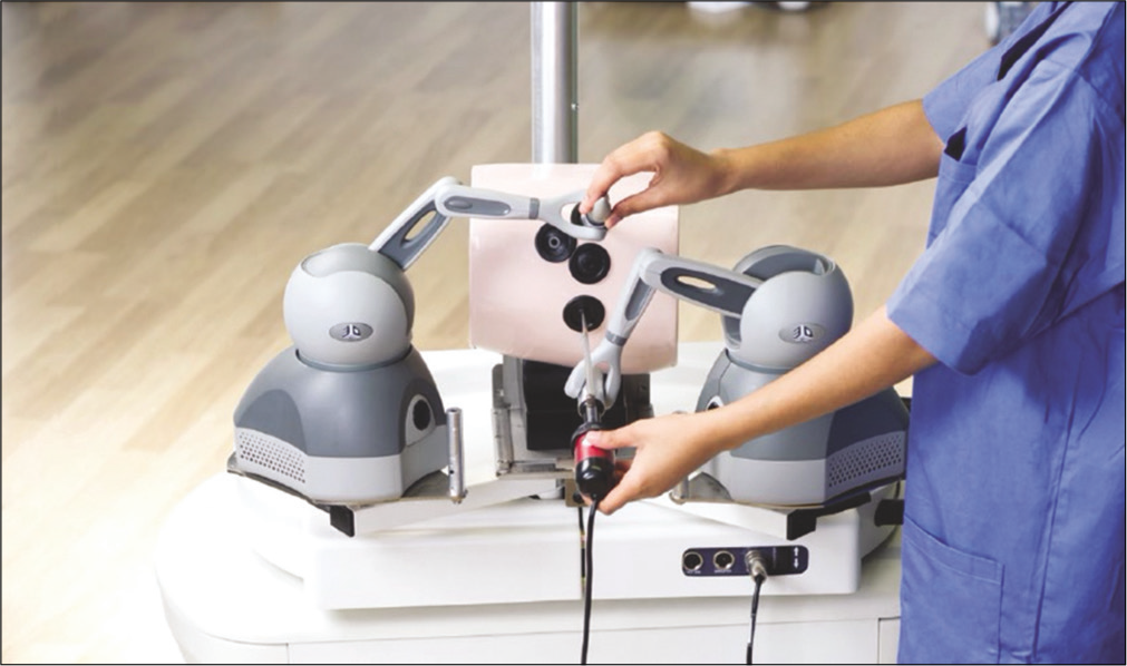
- Simbionix Arthro Mentro hip arthroscopy simulator.

- Visual examination (top left), basic probe examination (top right, bottom) using the Simbionix Arthro Mentor hip arthroscopy simulator.
Despite this, evidence of real-world benefit in HA is currently lacking. However, VR models of shoulder and knee arthroscopy confer a real-world benefit. Orthopedic trainees who receive VR simulator training outperform control trainees in a theater setting.[37] This, along with the recent validation of another novel VR simulator (VirtaMed ArthroS), may signal the beginning of VR simulation-based training for HA.[38] Indeed, VR simulation has already been adopted as a potential approach to simulation in the British Orthopaedic Association Trauma and Orthopaedic Training curriculum.[7]
Computer-assisted surgery
Computer-assisted technology in the orthopedic theater has enabled the emergence of patient-specific surgery. Computer-assisted surgery can be broadly thought of as a two-phase process, where there is computer-aided pre-operative assessment and subsequent intraoperative navigation.[39] In the field of HA, this is most applicable to the treatment of FAI. FAI presents in three forms (1) cam, where the head-neck offset is reduced, (2) pincer, where there is acetabular overcoverage, or (3) mixed type with features of both cam and pincer.[5] HA has been used to treat camdeformities and smaller pincer deformities. Larger pincer deformities often require open surgical procedures. While traditional arthroscopic treatment of FAI produces favorable patient-reported outcome measures (PROMs), arthroscopic resection of cam lesions results in a higher post-operative alpha angle than open arthrotomy, which represents an intrinsic limitation of HA.[40,41] The surgeon must assess the adequacy of bony resection during osteochondroplasty through the arthroscope and by fluoroscopy, which does not fully represent the 3D nature of the osseous deformity. Thus, a cam deformity may extend further anteriorly or posteriorly than the plane in which fluoroscopy shows that an adequate head-neck offset has been achieved.
Pre-operative computer-aided assessment enables surgeons to develop a precise operative plan. It affords visualization of a patient-specific reconstruction of the osseous anatomy based on prior radiographic investigations such as a 3D CT and enables accurate assessment of the extent of an impinging cam lesion. The surgeon can then develop a pre-operative plan for osteochondroplasty specific to this individual to resect only the amount of bone which would have resulted in impingement.[39] Current computer-assisted models have been shown to find higher alpha angles when compared to either XR or CT; these traditional assessment measures do not assess the cam deformity at its maximal extent which tends to be further anterosuperior.[42]
Computer-aided assessment also circumvents intrinsic problems with the alpha angle metric. The alpha angle does not take into account the extent of the head-neck offset in 3D, or the extent of the cam deformity, and does not always correlate with clinical range of motion (ROM).[43] The dependence on alpha angle prevents the development of a personalized management plan, in which the surgeon can accurately predict whether the patient will gain a normal pain-free ROM. Pre- operative assessment tools model impingement through either applied algorithms or virtual 3D reconstruction of hip anatomy, identifying the impinging site, and subsequently using impingement modeling to determine ROM measures. Such programs have been validated in cadaveric models and shown to predict an accurate ROM based on CT data.[44] These programs can then be used to evaluate surgical plans and predict the volume of resection required to achieve a normal non-impinging ROM.[45]
To achieve the benefits of pre-operative planning, navigation systems guide the surgeon to precisely reproduce these pre-operative plans intraoperatively.[39] These systems trace the surgical tool and match surgeon-guided movements to the pre-operative plan, thus tracking the intraoperative situation. This is achieved through an encoder linkage system which tracks surgical instruments during HA and was first developed by Monahan and Shimada in 2006.[46] For accurate tracking, there must be registration of the surgical equipment to the navigation system, producing correspondence between the pre-operative model and the intraoperative anatomy. Registration is achieved using fluoroscopy, or an imageless technique requiring a digitalized pointer to register bony landmarks. Evidence from a cadaveric model suggests that imageless registration techniques provide inaccurate navigation for HA, while fluoroscopic registration is sufficiently accurate for femoral osteochondroplasty.[47]
Clinical evidence into the efficacy of navigated FAI correction is conflicting and is further confounded by the use of different planning and navigation systems. One study published in 2009 showed that the use of a navigation system did not improve the rate of insufficient alpha angle correction in patients undergoing arthroscopic FAI correction.[48] Conversely, a randomized controlled trial published in 2017 showed a significant improvement in alpha angle for those patients who underwent navigated arthroscopic cam resection compared to conventional arthroscopy [Figures 4 and 5].[49] Larger studies are required to validate these positive findings. There is evidence that the adoption of navigated surgery may reduce the learning curve for HA among novice surgeons.[50] Almoussa et al. reported the same shaping accuracy of the femur to be achieved by both an experienced and novice surgeon when using navigated surgical techniques for the treatment of a cam-type FAI model.

- Analysis of simulated bony range of motion using Articulis software package (Clinical Graphics, Delft, The Netherlands) and suggested pre-operative resection plan on the femoral neck to normalize the range of motion defects. Reproduced from: “Accuracy of navigated cam resection in femoroacetabular impingement: A randomized controlled trial” by Van Houcke et al. reproduced with permission from John Wiley and Sons.

- The femoral marker (a) and fluoroscopy (b) are calibrated using the rigid pointer. An intra-operative fluoroscopy scan limited to the proximal femur is performed (c) to allow for image-based matching of the pre-operative plan. Finally, live resection control in relation to the pre-operative plan can be performed using the rigid pointer and fluoroscopy is no longer required (d). Reproduced from: “Accuracy of navigated cam resection in femoroacetabular impingement: A randomized controlled trial” by Van Houcke et al. reproduced with permission from John Wiley and Sons.
With such developments in computer-assisted surgery, it is hard not to question whether there will be a future role for robotic surgery in HA. Enaction of a quantitative computer- assisted pre-operative plan by automated surgical action would ensure that there is no deviation from a patient’s personalized plan. The adoption of robotic surgery in other specialties including laparoscopic surgery has led to improved outcomes for patients in specific procedures such as distal gastrectomy.[51] While robotic surgery is currently only feasible in a cadaveric model, it is assumed that this system will be amenable to HA due to the similarity of laparoscopic and arthroscopic instruments.[39]
Stratification of disease
Given the use of computer-aided pre-operative planning, and the controversies surrounding traditional disease measures, such as the alpha angle, it is possible that we will see a new era of disease stratification based on 3D pathoanatomic features. This strategy relies on good modeling of the equivalent normal anatomy for each patient and will provide further accurate classification of disease to improve the accuracy of the pre- operative planning process. Models for identifying and differentiating patient-specific anatomical variation of the femoral head-neck junction from pathological deviations of the osseous anatomy have already been developed and offer increased individual precision versus measures such as the alpha angle which are defined based on population averages.
Khanduja et al. used statistical shape models (SSM) to accurately describe the anatomy of the proximal femur and its variation using 10 shape variation inputs derived from CT data, predicting femoral surface anatomy with a small error [Figure 6].[52] When applied to both a control and patient group with cam-type FAI, the differences between alpha angle of the SSM model and the control group were non-significant, while in the patient group, a significant difference was detected in the 1 and 2 o’clock positions where the cam deformity is typically found. The quantification of this difference provides a comparison of the pathological cam deformity in the individual to the predicted normal anatomy of the proximal femur in this individual. Therefore, the deformity can be stratified by true severity, and pre-operative assessment of the resection volume required to attain normal anatomy for an individual can occur.

- Statistical shape modeling of proximal femur showing cam deformity by comparing actual and predicted morphology of the head-neck junction in individual patients. Actual morphology (a), predicted morphology (b), original scan (c) and virtual twin (d), reproduced from: “Patient-specific assessment of dysmorphism of the femoral head-neck junction: A statistical shape model approach”. Khanduja et al. Int J Med Robot. 2016;12(4):765-772. reproduced with permission from John Wiley and Sons.
Virtually modeling hip pathology will enable better classification of pathology through a thorough understanding of etiology. This is exemplified in a recent paper exploring the pathogenesis of internal snapping of the psoas tendon. Using SSM, the authors generated a model of the lower limb to investigate psoas tendon behavior.[53] This revealed the exact location of psoas tendon movement and identified underlying torsional femoral dysplasia as an associated anatomic risk factor in snapping hip syndrome. If this is to be confirmed clinically, it will inform a change in surgical treatment from current practice.
Further studies of this nature will enable accurate classification of disease and promote changes in surgical practice to better match the underlying pathology. It may also become possible to use these technologies in conjunction with imaging to inform differential diagnoses and enhance diagnostic capability.
Outcome-based intervention
The future of personalized surgical management of patients using computer-assisted surgical technology is bright, however, patient outcomes are multifactorial and do not only rely on a technically successful operation. In a recent systematic review, other risk factors affecting the outcome of FAI correction were patient characteristics and pre-operative PROMs.[41] Following this logic, a risk prediction model has been produced to predict the functional outcome of HA for FAI correction. This identified several pre-operative risk factors: (1) gender, indication: (2) pincer and (3) labral tear, along with pre-operative PROM scores including measures of psychological health.[54] Without taking into account intra-operative parameters, this model successfully predicted outcome in 71% of cases.
This demonstrates the importance of optimizing patient- specific risk factors to achieve the best possible outcomes. While some risk factors including female gender and pincer impingement cannot be modified, PROMs could potentially be improved preoperatively. For instance, one may question whether attempting to improve mental health scores preoperatively would result in improved outcomes. Not only would this improve psychological PROMs but also research suggests that the pre-operative symptoms of FAI are more related to a patient’s mental health scores than the pathomorphology within the joint.[55]
Given that, pre-operative PROMs can largely predict whether a successful outcome will occur, improving pre-operative PROMS may be a promising future avenue for personalized HA. Pre-operative physiotherapy may also be employed to optimize pre-operative PROMs. A small pilot study has shown that in patients undergoing HA for FAI, it may be possible for them to improve their pain, function, and muscle power postoperatively using a pre-habilitation program.[56] In addition, the senior author is investigating whether pre- habilitation has a role in patients undergoing orthopedic surgery.[57]
CONCLUSION
HA is a widely accepted treatment for a variety of hip conditions. Adequate disease stratification and assessment of pre-operative risk factors influence the long-term outcome following the HA.
Technological innovations and advances in biological therapies have enabled surgeons to increase the scope of HA and the future is certainly exciting and bright.
Declaration of patient consent
Patient’s consent not required as patients identity is not disclosed or compromised.
Financial support and sponsorship
Nil.
Conflicts of interest
Dr. Vikas Khanduja is on the Editorial Board of the Journal.
References
- Current concepts in the diagnosis and management of femoroacetabular impingement. Int Orthop. 2011;35:1427-35.
- [CrossRef] [PubMed] [Google Scholar]
- Hip arthroscopy versus best conservative care for the treatment of femoroacetabular impingement syndrome (UK FASHIoN): A multicentre randomised controlled trial. Lancet (London England). 2018;391:2225-35.
- [CrossRef] [Google Scholar]
- Historical review of arthroscopic surgery of the hip. Int Orthop. 2017;41:1983-94.
- [CrossRef] [PubMed] [Google Scholar]
- Age-related trends in hip arthroscopy: A large cross-sectional analysis. Arthroscopy. 2015;31:2307-13.
- [CrossRef] [PubMed] [Google Scholar]
- Femoroacetabular impingement: A cause for osteoarthritis of the hip. Clin Orthop Relat Res. 2003;417:112.
- [Google Scholar]
- Hip arthroscopy: Analysis of a single surgeon's learning experience. J Bone Joint Surg Am. 2011;93(Suppl 2):52-6.
- [CrossRef] [PubMed] [Google Scholar]
- Training young adult hip surgeons for the future: The Cambridge vision. Bone Joint 360. 2016;5:8-12.
- [CrossRef] [Google Scholar]
- Complications following arthroscopic surgery of the hip: A systematic review of 36 761 cases. Bone Joint J. 2017;99:1577-83.
- [CrossRef] [PubMed] [Google Scholar]
- Complications in hip arthroscopy. Muscles Ligaments Tendons J. 2016;6:402-9.
- [CrossRef] [PubMed] [Google Scholar]
- The impact of acetabular labral tears on sexual activity in women. J Hip Preserv Surg. 2019;6:301-3.
- [CrossRef] [PubMed] [Google Scholar]
- Genitoperineal injuries associated with the use of an orthopedic table with a perineal posttraction. J Trauma. 2008;65:820-3.
- [CrossRef] [PubMed] [Google Scholar]
- Complications of arthroscopic surgery of the hip. Bone Joint Res. 2012;1:131-44.
- [CrossRef] [PubMed] [Google Scholar]
- Hip arthroscopy distraction without the use of a perineal post: Prospective study. Orthopedics. 2013;36:e1-5.
- [CrossRef] [PubMed] [Google Scholar]
- Hip distraction without a perineal post: A prospective study of 1000 hip arthroscopy cases. Am J Sports Med. 2018;46:632-41.
- [CrossRef] [PubMed] [Google Scholar]
- Achieving post-free distraction in hip arthroscopy with a pink pad patient positioning device using standard hip distraction tables. Arthrosc Tech. 2019;8:e363-8.
- [CrossRef] [PubMed] [Google Scholar]
- The tutankhamun technique in hip arthroscopy. Arthrosc Tech. 2018;7:e1167-71.
- [CrossRef] [PubMed] [Google Scholar]
- Arthroscopic hip labral augmentation technique with iliotibial band graft. Arthrosc Tech. 2017;6:e351-6.
- [CrossRef] [PubMed] [Google Scholar]
- Arthroscopic acetabular labral repair using the Q-FIX suture anchor. Arthrosc Tech. 2019;8:e801-5.
- [CrossRef] [PubMed] [Google Scholar]
- Arthroscopic labral reconstruction with gracilis autograft. Arthrosc Tech. 2012;1:e15-21.
- [CrossRef] [PubMed] [Google Scholar]
- Labral reconstruction using the ligamentum teres capitis: Report of a new technique. Clin Orthop Relat Res. 2009;467:753-9.
- [CrossRef] [PubMed] [Google Scholar]
- Arthroscopic labral reconstruction in the hip using iliotibial band autograft: Technique and early outcomes. Arthroscopy. 2010;26:750-6.
- [CrossRef] [PubMed] [Google Scholar]
- Arthroscopic labralization of the hip: An alternative to labral reconstruction. Arthrosc Tech. 2014;3:e131-3.
- [CrossRef] [PubMed] [Google Scholar]
- The role of the ligamentum teres in the adult hip: Redundant or relevant? A review. J Hip Preserv Surg. 2018;5:15-22.
- [CrossRef] [PubMed] [Google Scholar]
- Arthroscopic reconstruction of the ligamentum teres. Arthroscopy. 2011;27:436-41.
- [CrossRef] [PubMed] [Google Scholar]
- Arthroscopic reconstruction of the ligamentum teres: Technique and early outcomes. J Bone Joint Surg Br. 2012;94:1494-8.
- [CrossRef] [PubMed] [Google Scholar]
- Ligamentum teres injuries of the hip: A systematic review examining surgical indications, treatment options, and outcomes. Arthroscopy. 2014;30:1634-41.
- [CrossRef] [PubMed] [Google Scholar]
- Arthroscopic ligamentum teres reconstruction using anterior tibialis allograft and the tension-slide technique. Arthrosc Tech. 2019;8:e1075-83.
- [CrossRef] [PubMed] [Google Scholar]
- Outcomes of cartilage repair techniques for chondral injury in the hip-a systematic review. Int Orthop. 2018;42:2309-22.
- [CrossRef] [PubMed] [Google Scholar]
- Treatment of deep cartilage defects in the knee with autologous chondrocyte transplantation. N Engl J Med. 1994;331:889-95.
- [CrossRef] [PubMed] [Google Scholar]
- Five-year results of arthroscopic techniques for the treatment of acetabular chondral lesions in femoroacetabular impingement. Int Orthop. 2014;38:2057-64.
- [CrossRef] [PubMed] [Google Scholar]
- Feasibility of arthroscopic placement of autologous matrix-induced chondrogenesis grafts in the cadaver hip joint. Orthop Rev (Pavia). 2013;5:26.
- [CrossRef] [PubMed] [Google Scholar]
- Injectable autologous chondrocyte implantation (ACI) in acetabular cartilage defects-three-year results. J Hip Preserv Surg. 2018;5:386-92.
- [CrossRef] [PubMed] [Google Scholar]
- The learning curve for hip arthroscopy: A systematic review. Arthroscopy. 2014;30:389-97.
- [CrossRef] [PubMed] [Google Scholar]
- The role of simulation in developing surgical skills. Curr Rev Musculoskelet Med. 2014;7:155-60.
- [CrossRef] [PubMed] [Google Scholar]
- Prospective, randomized assessment of transfer of training (ToT) and transfer effectiveness ratio (TER) of virtual reality simulation training for laparoscopic skill acquisition. Ann Surg. 2013;257:1025-31.
- [CrossRef] [PubMed] [Google Scholar]
- The learning curves of a validated virtual reality hip arthroscopy simulator. Arch Orthop Trauma Surg. 2020;140:761-7.
- [CrossRef] [PubMed] [Google Scholar]
- Does virtual reality simulation have a role in training trauma and orthopaedic surgeons? Bone Joint J. 2018;100B:559-65.
- [CrossRef] [PubMed] [Google Scholar]
- Validation of a novel hip arthroscopy simulator: Establishing construct validity. J Hip Preserv Surg. 2019;6:385-9.
- [CrossRef] [PubMed] [Google Scholar]
- Review: Current concepts in computer-assisted hip arthroscopy. Int J Med Robot Comput Assist Surg. 2018;14:1-8.
- [CrossRef] [PubMed] [Google Scholar]
- Hip arthroscopy versus open surgical dislocation for femoroacetabular impingement: A systematic review and meta-analysis. Medicine (Baltimore). 2016;95:e5122.
- [CrossRef] [PubMed] [Google Scholar]
- Systematic review and meta-analysis of outcomes after hip arthroscopy in femoroacetabular impingement. Am J Sports Med. 2019;47:488-500.
- [CrossRef] [PubMed] [Google Scholar]
- Novel CT-based three-dimensional software improves the characterization of cam morphology. Clin Orthop Relat Res. 2013;471:2484-91.
- [CrossRef] [PubMed] [Google Scholar]
- Clinical measures of hip range of motion do not correlate with the degree of cam morphology in semi-elite australian footballers: A cross-sectional study. Int J Sports Phys Ther. 2017;12:1078-86.
- [CrossRef] [PubMed] [Google Scholar]
- Noninvasive three-dimensional assessment of femoroacetabular impingement. J Orthop Res. 2007;25:122-31.
- [CrossRef] [PubMed] [Google Scholar]
- Range of motion in anterior femoroacetabular impingement. Clin Orthop Relat Res. 2007;458:117-24.
- [CrossRef] [PubMed] [Google Scholar]
- Computer-aided navigation for arthroscopic hip surgery using encoder linkages for position tracking. Int J Med Robot. 2006;2:271-8.
- [CrossRef] [PubMed] [Google Scholar]
- Imageless versus image-based registration in navigated arthroscopy of the hip. J Bone Joint Surg Br. 2012;94:624-9.
- [CrossRef] [PubMed] [Google Scholar]
- Evaluation of a computed tomography-based navigation system prototype for hip arthroscopy in the treatment of femoroacetabular cam impingement. Arthroscopy. 2009;25:382-91.
- [CrossRef] [PubMed] [Google Scholar]
- Accuracy of navigated cam resection in femoroacetabular impingement: A randomised controlled trial. Int J Med Robot Comput Assist Surg. 2017;13:1-7.
- [CrossRef] [PubMed] [Google Scholar]
- Computer-assisted correction of cam-type femoroacetabular impingement: A Sawbones study. J Bone Joint Surg Am. 2011;93(Suppl 2):70-5.
- [CrossRef] [PubMed] [Google Scholar]
- Comparison of surgical outcomes between integrated robotic and conventional laparoscopic surgery for distal gastrectomy: A propensity score matching analysis. Sci Rep. 2020;10:1-9.
- [CrossRef] [PubMed] [Google Scholar]
- Patient-specific assessment of dysmorphism of the femoral head-neck junction: A statistical shape model approach. Int J Med Robot Comput Assist Surg. 2016;12:765-72.
- [CrossRef] [PubMed] [Google Scholar]
- Mechanics of psoas tendon snapping. A virtual population study. Front Bioeng Biotechnol. 2020;8:1-13.
- [CrossRef] [PubMed] [Google Scholar]
- Developing a risk prediction model for the functional outcome after hip arthroscopy. BMC Musculoskelet Disord. 2018;19:122.
- [CrossRef] [PubMed] [Google Scholar]
- Preoperative symptoms in femoroacetabular impingement patients are more related to mental health scores than the severity of labral tear or magnitude of bony deformity. J Arthroplasty. 2017;32:3603-6.
- [CrossRef] [PubMed] [Google Scholar]
- The HAPI hip arthroscopy pre-habilitation intervention' study: Does pre-habilitation affect outcomes in patients undergoing hip arthroscopy for femoro-acetabular impingement? J Hip Preserv Surg. 2017;4:hnw046.
- [CrossRef] [PubMed] [Google Scholar]
- Effectiveness of prehabilitation for patients undergoing orthopaedic surgery: Protocol for a systematic review and meta-analysis. BMJ Open. 2019;9:e031119.
- [CrossRef] [PubMed] [Google Scholar]


