Translate this page into:
History of arthroscopy in India: Origins and evolution

*Corresponding author: David V. Rajan, MS Ortho, MNAMS, FRCS (G), Senior Consultant Orthopaedic Surgeon, Ortho One – Orthopaedic Speciality Centre, 657, Trichy Road, Coimbatore, Tamil Nadu, India. davidvrajan@gmail.com
-
Received: ,
Accepted: ,
How to cite this article: Rajan DV, Ashraf M, Challumuri N, Sahanand SK. History of arthroscopy in India: Origins and evolution. J Arthrosc Surg Sport Med 2020;1(1):5-10.
Abstract
The practice of arthroscopy in India had started as early as 1978; and during the same year, the Indian chapter of the International Arthroscopy Association was drafted alongside other countries such as Australia and Brazil. The subspecialty of arthroscopy has been a boon to both; the orthopedic surgeon and the patient. The advent of arthroscopy has enabled the orthopedic surgeon to clearly visualize and delineate the extent of disease, with minimal invasion. Moreover, the patient is benefited with rapid recovery and an early return to activities. The present-day arthroscopic surgeries include diagnostic arthroscopy, ligament reconstruction, cartilage repair, and labral repairs and have undoubtedly evolved into a glamorous subspecialty in orthopedics. However, before the technological advancements, the technique of arthroscopy had modest origins. This review traverses through the history of arthroscopy with special emphasis on the advances of arthroscopy in India.
Keywords
History
Arthroscope
Pioneers
India
Minimally invasive
Reconstruction
Advancements
INTRODUCTION
The practice of arthroscopy in India had blossomed almost at the same time as that of other developed nations. However, the progress in the earlier days was not as rapid as was expected. The reason for this slow progress was multifactorial and could be attributed to the difficulty in organizing and setting up an arthroscopy unit, a steep learning curve for surgeons, reduced monetary benefits, expensive equipment, and the socioeconomic status of our country at that time. The arthroscope was initially developed to visualize the diseased knee joint. The direct viewing arthroscope was eventually replaced by the modern-day arthroscope. Thereafter, the utility has expanded to treating knee injuries with meniscectomies and synovial pathologies. Prof Masaki Watanabe, the father of modern arthroscopy, had described the growth of arthroscopy in four stages. The first stage was from 1920 to 1959, when various designs of arthroscopes were being made and experimented on. The second stage was from 1960 to 1969 and marked the practical application of arthroscopy to diagnose and treat diseased knees. The third stage was from 1970 to 1978, in which the first commercially available 21 no. arthroscope was introduced in countries such as the Unites States, Canada, France, and England. The fourth stage was from 1979 onwards, and in this phase, the incorporation of cold light replaced incandescent light. The use of the arthroscope into other joints such as shoulder, hip, and ankle was performed during this phase.[1]
In India, although arthroscopy started earlier, it did not really catch up with orthopedic surgeons. On the other hand, the early 90’s saw the rise of the use of the laparoscope by general surgeons and laparoscopy grew more rapidly than the use of the arthroscope by orthopedic surgeons. This disparity was mostly due to the steep learning curve associated with arthroscopy and also the emergence of other subspecialties such as arthroplasty, trauma, and internal fixation which was more feasible for the orthopedic surgeon. This review highlights the history and development of arthroscopy with an Indian perspective.
ANCIENT HISTORY OF SURGERY IN INDIA
The earliest documented evidence of surgical procedures and surgical instruments were reported in the compendium titled “Sushruta Samahita” compiled by Susruta. He belonged to a period between 600 and 800 BC. Susruta’s works are renowned world over for the surgical procedures he had practiced during that period. However, little is known about his descriptions on surgical instruments and endoscopes in particular. He had developed several surgical instruments which were categorized into sharp instruments and blunt instruments. Of which, blunt tubular instruments were called as the “Nadi Yantra.” The “Nadi Yantra” is similar to the modern-day endoscope which is used to examine body cavities and external orifices. This is a useful tool which aids in inspection and treatment. The inquisitiveness of Susruta to look into cavities for diagnosis was way ahead of his time.[2]
WORLD HISTORY OF ARTHROSCOPY
Before the advent of arthroscopy, the efforts to view closed cavities were always of prime importance to surgeon’s world over. It was in 1806 that Bozzini reported his first attempt to view the bladder using instrument called as “Lichtletr.” It was not until 1886, the first cystoscope with illumination was developed by Leiter and Nitze in Germany. Following this the laparo-thoracoscope was invented in 1910 by Hans Christian Jacobeus, a Swedish Physician. With subsequent improvement in lens optical systems, these were incorporated into the endoscope for better viewing.
The use of endoscope in joints was known as “Arthro- endoscope,” however, this practice started after the successful use of the cystoscope and laparothoracoscope. The first reported contribution on arthroscope was by Severin Nordentoft in 1912. He had used a 5 mm diameter trocar similar in design to the Jacobeus laparothoracoscope.
However, the true inventor and developer of arthroscopy are credited to Kenji Takagi from Tokyo, Japan. His works were largely related to tubercular knees and that early diagnosis would lead to better treatment and prevent ankylosis. His first arthroscope, developed in 1920, was 7.3 mm and was found to be not suitable for clinical practice. Over the years, he worked on various designs and modifications.
By 1931 developed his first arthroscope which was a 3.5 mm diameter trocar. During this period, he had also emphasized the importance of using normal saline for distention of the knee joint for better visualization. And by 1938, he had developed his 12th design of the arthroscope.
After Kenji Takagi, it was his prodigy Masaki Watnabe who continued the work from where his mentor left. With the help of evolving techniques in light and optics, he was able to design and develop several arthroscopes. Eventually in 1958, the 21st design also known as the 21st arthroscope was developed with a 6 mm sheath and had magnificent lens with a field of vision of 101 degrees and a depth focus from 1 mm to infinity. The 21st arthroscope became the world’s first commercially produced arthroscope. It was also the last scope to use an incandescent light source.
In 1970, the 25th arthroscope was developed, it contained an ultrathin fiber-optic which had a 2 mm diameter sheath and a single “selfoc” fiber (1.7 mm in diameter) to transmit to the eye.[3-5]
During this period, arthroscopy was performed through direct vision through the telescope. For recording purposes, the images from the eye piece were captured by an SLR camera which was connected to the eye piece by an adapter. This is how the early atlas of arthroscopy was published.
Once the video camera was developed, it enabled the surgeon to look at the monitor and perform the surgery. Although the camera was bulky, it was a great boon to the surgeon and the team. Everyone could participate in the surgery by looking at the image on the monitor. However, the cost of procuring this equipment was prohibitive for the Indian surgeon. As the saying goes “necessity is the mother of all inventions,” Indian arthroscopists developed an indigenous alternative to this by connecting the surveillance camera to view the image in the monitor. Although the clarity was compromised, some surgeons were using this indigenous way for cost efficiency.
For better clarity and visualization of the surgical field, irrigation systems are imperative. Dr. Kenji Takagi was the first to use normal saline as the irrigation fluid. Dr. Eugen Bircher was credited to using CO2 insufflation for dry scopy. The fluid management systems have evolved and various generations of pump systems have been developed. Gravity flow systems were used first and are still commonly used followed by automated pump systems. Gravity flow systems simply use gravity to control inflow by positioning a bag of fluid higher than the joint to provide enough pressure for insufflation. The development and use of an automated pump for arthroscopy began in Sweden in the 1970s. At present, there are two basic types of automated pump systems. The first type is a pressure control pump, which controls pressure through inflow only. More recent automated pump systems maintain pressure by controlling inflow and outflow independently, thus termed pressure and flow control pumps or dual systems.[6]
Ever since these pioneering works had been brought to daylight, there have been huge strides in the advancement of arthroscopy as a specialty. A brief summary of these advancements is outlined in [Table 1].
| 1912 | Severin Nordentoft, first to use endoscope in knees |
| 1918 | Kenji Takagi treats tuberculous knees with scope |
| 1921 | Eugen Bircher credited to using CO2insufflation for dry scopy |
| 1954 | Harold Hopkins develops glass fiber cold light |
| 1955 | Masaki Watanabe, father of modern arthroscopy, develops concept of triangulation |
| 1959 | The Watanabe No. 21 arthroscope, commercial production |
| 1962 | Masaki Watanabe performs first arthroscopic meniscectomy |
| 1967 | The Watanabe No.22 arthroscope, first to use cold light |
| 1968 | Robert Jackson, first instructional course on arthroscopy at the AAOS |
| 1974 | IAA is founded |
| 1978 | Indian chapter of IAA |
| 1982 | Arthroscopy Association of North America is founded |
| 1983 | Indian Arthroscopy Society is founded by Dr. Pravin Kanabar, Dr. Pravin Vohra, and Shahthe |
| 1984 | European Society of Sports Traumatology, Knee Surgery and Arthroscopy is founded |
| 1995 | First Shoulder Arthroscopy Live Surgical Demonstration in India, held at Coimbatore |
| 1996 | Shoulder Society of India was founded by Dr. Devadoss and Dr. David Rajan |
| 2002 | First Annual Indian Arthroscopy Society conference heldat Coimbatore |
| 2012 | Shoulder Society of India merged to form Shoulder and Elbow Society of India |
IAA: International Arthroscopy Association
Early introduction of arthroscopy in India
At a time, when the principles and teachings for fracture fixations by AO trauma were prevalent, arthroscopy too had a foothold in India. During this period, the practice of orthopedics in India was predominantly focused on fracture fixations, deformity correction, soft-tissue procedures, and replacement surgeries. Arthroscopy remained a surgical exercise for those who were passionate about it and for those who could find time after their practice of trauma. Barely two decades after the first arthroscopic meniscectomy by Watanabe, the Indian chapter of the International Arthroscopy Association (IAA) was founded. A few years later, in 1983, the Indian Arthroscopy Society was formed.
During these formative years, the monumental efforts of Dr. Pravin Kanabar (Ahmedabad), Dr. Pravin Vohra (Bombay), and Dr. Sathe (Bombay) are to be mentioned as they were instrumental in the formation of the organization and conduct of instructional courses. They had a great passion for arthroscopy which led them to purchase their own equipment which costs a fortune and started their practice with minimal formal training in the field. In 1978, the trio was the first to perform arthroscopy in India at their respective centers. With the zero-degree viewing scope, Dr. Pravin Kanabar was quoted stating that “All he could see was the anterior horn of medial meniscus and it was a Joyous moment.” In the following years, surgeons from all over the world had descended to India and conducted instructional courses which would go on to enthuse hundreds of surgeons to take up arthroscopy.
Phases of arthroscopy in India
Phase I “The period of awareness” [1985–1989]
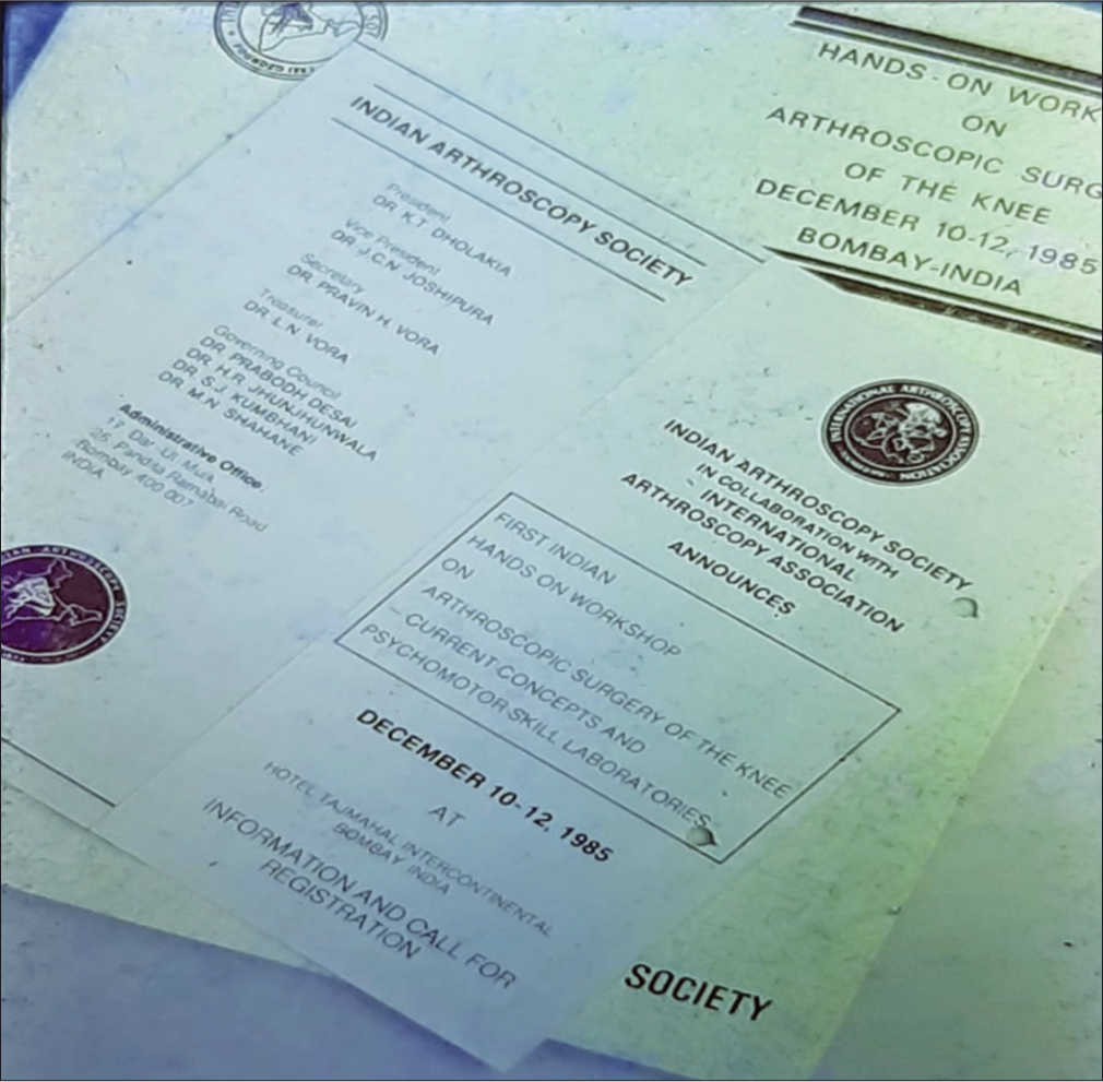
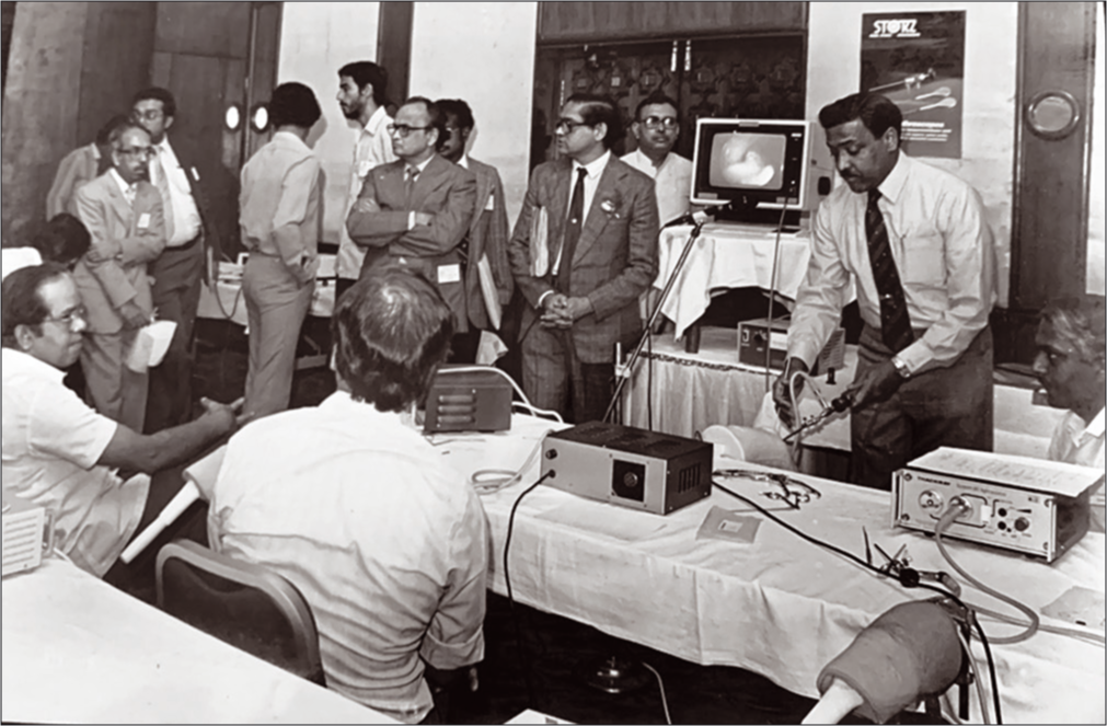
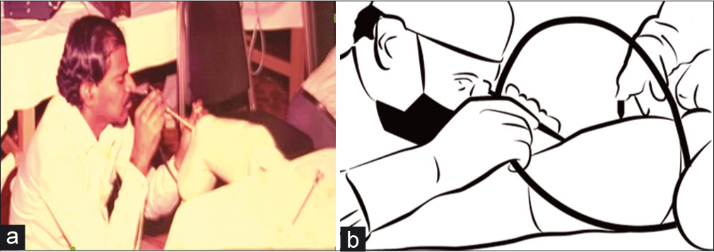
After the formation of the arthroscopy society and conduct of regular meetings on arthroscopy, there was an awakening among orthopedic surgeons about a new subspecialty which was being practiced world over. In 1983, a series of informal lectures/demonstrations of the direct viewing arthroscope was held by Dr. Dinesh Patel (USA) in Ahmedabad, Dr. Gopalakrishnan (USA) in Bengaluru, and Dr. Harold Eikler (Holland) in Coimbatore. This had enthused the minds of orthopedic surgeons and lead to the 1st international arthroscopy meeting in India held in Bombay, 1985 [Figure 1]. The meeting was the first hands on instructional course chaired by Dr. Dinesh Patel (USA) and Dr. Gopalakrishnan (USA). The meeting was eventually called the “Big Bombay meeting” as it had over 500 delegates and the faculty included Dr. Bob Jackson (USA), Dr. Cerulli (Germany), Dr. Witvity (Germany), and others. Subsequent meetings were held in 1988 at Madras and in 1989 at Ahmedabad [Figures 2 and 3(a,b)]. Furthermore, the likes of Dr. Ramon Cugat (Spain) and Dr. Jaap Willems (Holland) had frequent visits to different places in India and held live demonstrations.[7]

- Brochure sent to orthopedic surgeons in India, inviting them for the first ever formal arthroscopy meeting organized by International Arthroscopy Association at Bombay in 1985.

- Dr. Gopalkrishnan demonstrating knee arthroscopy at an instructional course in 1988 in Madras. In the background is Dr. Sriram and Dr. Vora.

- (a) 1988 Chennai workshop hands on sessions on model. (b) Diagrammatic representation of surgery being performed by direct viewing.
Due to the expenses associated with procuring instruments, few independent surgeons would take the journey overseas (USA and Europe) to purchase the instruments, such was the euphoria associated with arthroscopy. Following the plethora of meetings and demonstrations, there was an assumption among the native surgeons to hold arthroscopy as a panacea for treating osteoarthritis with the use of scope and shaver. This practice, however, did not yield successful treatment outcomes and surgeons ended up in despair when in comparison with their arthroplasty counterparts. Adding to this, a steep learning curve and expensive equipment associated with arthroscopy had impeded the growth of arthroscopy after the initial spurt.
Phase II “Low Period” [1990–1998]
From the year 1990 to 1998, in India, trauma and arthroplasty seemed to put arthroscopy on the sidelines. The cost efficiency of arthroscopic surgery seemed to be less practical as the imported hardware was attracting custom duty of 100–200%. To understand this situation better, British arthroscopist, Dr. David Dandy in 1990 after one of his visits to India, had quoted saying that “There was a 200% import duty on electrical goods and optical items brought to India and if the financial position of Indian arthroscopists were applied to the U.S.A., the total cost of equipment for arthroscopic surgery would be approximately $l.5 million.” Such was the standard of policymaking which existed at that time.[8]
Obtaining arthroscopy equipment was considered unrealistic and remained a distant dream for many surgeons who wished to practice arthroscopy. Undeterred, some passionate surgeons who wanted to practice arthroscopy would hand carry the hardware on the way back from North America or Europe after attending meetings/courses.
Phase III “Period of Reawakening” [1999–2006]
However, things changed in the mid-90’s, when the period of liberalization had set in under the newly formed government. Instruments and implants were more easily readily available and it was possible for the surgeon as well as the industry to make their ends meet.
The struggles faced by the arthroscopists till about the late 90’s in setting up their unit were tremendous. It took about a decade after which specialists had emerged and they had trainees under them. This could possibly explain the deficit of contribution to literature by our arthroscopy surgeons up until the dawn of the new millennium. However, since then, there has been no looking back; there has been over a thousand articles published in the decade between 2006 and 2016 alone.
Phase IV “The Golden Era” 2007 onwards
The period from 2007 can be considered as “The Golden period” for practicing arthroscopic surgery. This can be attributed to the coherence which had developed between the surgeon, industry, and the society. Our society had become more affluent, awareness about health insurance was on the rise, a shift from motor vehicle accidents to sports injures was noted, and knowledge about minimally invasive surgery and its outcomes was prevalent.[9,10] The industry could develop ergonomic devices which suit the surgeon and also procurement of equipment was easier compared to earlier days. For the surgeon, he could begin to make his ends meet and setting up an arthroscopy unit was becoming cost efficient. Eventually, the surgeon could match with the expectations of the patient (predominantly a rising middle class). Furthermore, training initiatives had gained momentum in this period. Earlier many surgeons had to travel abroad for hands on cadaver training, as the saw bone model kind of training was the only available training in India. Nevertheless, the cadaver programs in SRMC, Chennai, and Ramaiah in Bengaluru gave a tremendous boost to the training of Indian arthroscopic surgeons. The companies such as Smith and Nephew, Arthrex, Stryker, and Storz have continued to strive and put in great effort in training the arthroscopy surgeon.
The emergence of shoulder arthroscopy
Shoulder arthroscopy was being routinely performed in the early 90’s at premier centers across the globe. However, it surfaced in India around the same time. The potential of this procedure was envisaged by a small group of surgeons and this led to the country’s first live arthroscopic shoulder surgery held in Coimbatore, 1995. The faculty included Dr. Jaap Willems (Netherlands) and a young surgeon from France with the name Dr. Pascal Boileau. However, there was a prevalence of suspicion about this new facet of arthroscopy in the minds of other orthopedic surgeons. However, the fascination grew into passion and thus was formed the Shoulder Society of India (SSI) in 1996 by Dr. Devadoss (Madurai) and Dr. David V. Rajan (Coimbatore) (Principal author). Ever since, there have been dedicated half-day specialty sessions exclusively for shoulder arthroscopy in the annual meetings of Indian Orthopaedic Association (IOA) [Figure 4].
![1992 IAS meeting during IOA annual meeting (From left to right: Dr. Pravin Vohra [Bombay], Dr. Nicholas Antao, Dr. Dinesh Patel [Boston, USA], Dr. Gopalkrishnan [Texas, USA], and Dr. SS Yadav [President IOA]).](/content/115/2020/1/1/img/JASSM-1-005-g004.png)
- 1992 IAS meeting during IOA annual meeting (From left to right: Dr. Pravin Vohra [Bombay], Dr. Nicholas Antao, Dr. Dinesh Patel [Boston, USA], Dr. Gopalkrishnan [Texas, USA], and Dr. SS Yadav [President IOA]).
Knee and shoulder arthroscopy were almost done exclusively by arthroscopic surgeons. Nowadays, limb-specific surgeons (upper limb surgeons and lower limb surgeons) have emerged and routinely perform arthroscopy, arthroplasty, and trauma pertaining to the particular extremity. To change with time, the SSI was eventually modified to form the Shoulder and Elbow Society of India in 2012.
Arthroscopy-related associations
The Indian Arthroscopy Society was formed in 1983 and has been active ever since its inception for the growth of arthroscopy in India. The IAS is chaired by the president who is elected on a term basis. The IAS has seen many doyens at its forefront who have made this association internationally acclaimed. At present (to date of this review), there are more than 2000 members associated with the IAS.
The International Society of Arthroscopy, Knee Surgery and Orthopaedic Sports medicine (ISAKOS) was formed in 1995 by the merger of the International Society of the Knee and IAA. Since then, ISAKOS has grown to a robust society of more than 3000 members representing more than 90 countries worldwide.[11]
The Arthroscopy Association of North America is an international professional organization of more than 5000 orthopedic surgeons and other medical professionals who are committed to advancing the field of minimally invasive orthopedic surgery to improve patient outcomes.[12]
Arthroscopy-related conferences
The annual meeting of IAS has been conducted regularly since the first annual meeting held in Coimbatore in 2002. The conference attracts a lot of passionate arthroscopy surgeons from India as well as international surgeons.
Numerous conferences in the specialty of shoulder arthroscopy and knee arthroscopy are conducted and these conferences usually have cadaver workshops which help the surgeons to enhance and replenish his anatomical knowledge.
The Pune knee course held annually gathers a large crowd each year as it serves as a basic course for beginners and a refresher course for the experts.
The cadaver courses for world class training are available at select centers such as MS Ramaiah Medical College, Bengaluru and Sri Ramachandra Medical University, Chennai.
Fellowship programs in arthroscopy
As in ancient days, the guru-shishya relationship was imperative for imbibing the professional and technical skills; similarly, the art of practicing arthroscopy was passed onto the successive generation by progressive minded mentors. Such mentorship has enabled the younger surgeons to expand their base and as a result the field of arthroscopy has grown to a great extent. The professional training is imparted by the various fellowship courses offered across various parts of our country. Fellowship programs are offered by the National Board of Examination, New Delhi, medical universities, orthopedic organizations, specialty centers, and also by individual mentors. The duration of fellowship varies from 6 months to a year. Furthermore, available is the short travelling fellowships which provide the fellows an insight into the field of arthroscopy. The “Train and Shine” initiative by the IAS matches the prospective fellow to an institute desirous of proving arthroscopy training. The IAS provides numerous fellowship opportunities each year such as the Dr. Gopalkrishnan fellowship, CASM fellowship, and CASM international travelling fellowship. The national board, New Delhi conducts the FNB courses in Sports medicine and arthroscopy. There are a few recognized centers for training by the ISAKOS in India.
Future of arthroscopy in India, the way to go
For any subspecialty or a surgical discipline to flourish you need technology, expertise and it should be affordable for the patient. In surgical disciplines in any country, minimally invasive techniques are replacing the conventional ones. For an arthroscopic surgery, the indications are ever expanding. Arthroscopic surgery is a quality of life surgery. Earlier in India, the acceptance for arthroscopic surgery was poor. The money in sports and for sporting activities was very little. With the introduction of various competitive sports such as ISL and IPL, there is now more money in sport and the athletes desire to get back to their original level of activity as quickly as possible in case of injury. With liberalization of the economy and improvement in quality of life of the common man, affordability for arthroscopy has improved. Certainly, most of the indigenous implants available now are of standard quality. Awareness about medical insurance and policies is another important factor for the increase in affordability of arthroscopic surgery. With better expertise and emergence of GP practice, the subspecialty has a great future. Efforts by the various societies and industries to conduct, cadaver training programs, etc., will definitely increase the demand for arthroscopic surgery. It is no surprise that the arthroscopic society which was started with around 40–45 members has now multiplied 50-fold till date.
Acknowledgments
The authors would like to acknowledge the inputs through personal communications from Dr. Gopalakrishnan, Dr. Pravin Kanabar, Dr. Sripathy Rao, Dr. Ashok Rajagopal, Dr. Anant Joshi, Dr. Nicholas Antao, Dr. Kanchan Bhattacharya, Dr. Vishnu Bhasin, and Dr. Sriram. Also included are instances recollected from the principal authors’ (DVR) experience of over three decades.
Declaration of patient consent
Patient’s consent not required as patients identity is not disclosed or compromised.
Financial support and sponsorship
Nil.
Conflicts of interest
Dr. S.K. Sahanand is on the Editorial Board of the Journal.
References
- Surgical instruments and endoscopes of Susruta, the sage surgeon of ancient India. Indian J Surg. 2008;70:219-23.
- [CrossRef] [PubMed] [Google Scholar]
- Arthroscopic surgery: A historical perspective. Orthop Nurs. 2008;27:349-54.
- [CrossRef] [PubMed] [Google Scholar]
- Use of an irrigation pump system in arthroscopic procedures. Orthopedics. 2016;39:e474-8.
- [CrossRef] [PubMed] [Google Scholar]
- Personal Communication through Email: haddidoctor1940@gmail.com on March 27
- Arthroscopy in other countries: Presidential guest lecture. Arthroscopy. 1990;6:248-51.
- [CrossRef] [Google Scholar]
- From the scalpel to the scope: The history of arthroscopy. In: Baylor University Medical Center Proceedings. Vol 9. United Kingdom: Taylor & Francis; 1996. p. :77-9.
- [CrossRef] [Google Scholar]
- The top 10 most cited Indian articles in arthroscopy in last 10 years. Indian J Orthop. 2017;51:505-15.
- [CrossRef] [PubMed] [Google Scholar]
- Evolution and current status of arthroscopic surgery in India. J Postgrad Med Educ Res. 2018;52:22-5.
- [CrossRef] [Google Scholar]
- Available from: https://www.isakos.com/Archives/Historical-Timeline [Last accessed on 2020 May 27]
- Available from: https://www.aana.org/AANAIMIS/Members/About%20AANA/Members/About-AANA/Overview [Last accessed on 2020 May 27]


